 Back
BackAxial Skeleton: Structure and Function of the Skull and Vertebral Column
Study Guide - Smart Notes
 Tailored notes based on your materials, expanded with key definitions, examples, and context.
Axial Skeleton Overview
Definition and Components
The axial skeleton forms the central axis of the human body, providing support and protection for the brain, spinal cord, and thoracic organs. It consists of the skull, vertebral column (including sacrum and coccyx), and the thoracic cage (ribs and sternum).
Axial skeleton: Skull, vertebral column, thoracic cage
Appendicular skeleton: Upper and lower limb bones
Classification of Bones by Shape
Types of Bones
Bones are classified based on their shapes, which relate to their functions and locations in the body.
Long bones: Longer than wide, with a shaft and two ends (e.g., humerus)
Short bones: Cube-shaped, found in wrist and ankle
Sesamoid bones: Develop within tendons (e.g., patella)
Flat bones: Thin, flattened, and curved (e.g., sternum, skull bones)
Irregular bones: Complex shapes (e.g., vertebrae, hip bones)
Sutural bones: Small bones within skull sutures (Wormian bones)
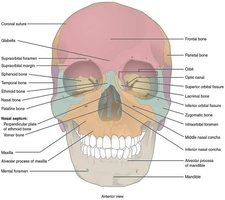
Skull Structure
Major Regions and Bones
The skull is composed of cranial and facial bones. The cranial bones protect the brain, while the facial bones form the structure of the face.
Cranial bones (8): Occipital, frontal, sphenoid, ethmoid, 2 parietal, 2 temporal
Facial bones (14): Maxilla (2), lacrimal (2), nasal (2), zygomatic (2), mandible, palatine (2), inferior nasal conchae (2), vomer
Sutures are immovable joints connecting cranial bones:
Sagittal suture
Lambdoid suture
Squamous suture
Coronal suture
Anterior View of the Skull
The anterior view displays the bones forming the forehead, orbits, nasal cavity, nasal septum, and jaws.
Key Skull Features
Hard palate: Formed by maxillary and palatine bones
TMJ (temporomandibular joint): Only movable joint of the face (mandible and temporal bone)
Teeth: Anchored in mandible and maxillary bones
Specialized Bones and Structures
Sphenoid bone: Forms part of cranial floor, contains sella turcica for pituitary gland, has multiple foramina for nerves and vessels
Ethmoid bone: Forms part of nasal septum, nasal cavity, and medial orbit wall; contains crista galli, perpendicular plate, cribriform plates, and ethmoid air cells
Nasal conchae: Superior and middle (ethmoid), inferior (independent); increase surface area and direct airflow in nasal cavity
Hyoid bone: Does not articulate with other bones; supports tongue and larynx muscles
Sinuses
Paranasal Sinuses
Sinuses are air-filled cavities within certain skull bones. They lighten the skull, add resonance to the voice, and drain into the nasal cavity.
Frontal sinus: Pain in forehead if infected
Ethmoid sinus: Pain between eyes if infected
Maxillary sinus: Pain in upper jaw if infected
Sphenoid sinus: Pain in back of eye if infected
Vertebral Column
Structure and Regions
The vertebral column supports the body, protects the spinal cord, and allows flexible movement. It consists of 24 vertebrae, the sacrum, and the coccyx.
Cervical vertebrae (C1–C7): Neck region
Thoracic vertebrae (T1–T12): Upper back, articulate with ribs
Lumbar vertebrae (L1–L5): Lower back, largest vertebrae
Sacrum: Fusion of 5 sacral vertebrae
Coccyx: Fusion of 4 small coccygeal vertebrae
Curvatures of the Spine
Primary curvatures: Thoracic and sacrococcygeal (present at birth)
Secondary curvatures: Cervical and lumbar (develop after birth)
Abnormal curvatures:
Scoliosis: Lateral bending
Kyphosis: Excessive thoracic curvature
Lordosis: Excessive lumbar curvature
Structure of a Typical Vertebra
Body: Weight-bearing portion
Vertebral arch: Formed by pedicles and laminae
Processes: Transverse, spinous, superior and inferior articular
Vertebral foramen: Passage for spinal cord
Intervertebral foramen: Passage for spinal nerves
Intervertebral discs: Fibrocartilage pads (anulus fibrosus and nucleus pulposus) between vertebral bodies
Special Vertebrae
Atlas (C1): Supports skull, allows "yes" motion
Axis (C2): Has dens, allows "no" motion
Atlas (C1) and axis (C2): "No" movement
L5 and sacrum, sacrum and coccyx, sacrum and ilium: Support and transfer of body weight
Thoracic Cage
The thoracic cage protects thoracic organs and supports respiration. It consists of the sternum, ribs, and thoracic vertebrae.
Sternum: Manubrium, body, xiphoid process
Ribs (12 pairs):
True ribs (1–7): Directly attached to sternum
False ribs (8–12): Indirect or no attachment to sternum
Floating ribs (11–12): No anterior attachment
Newborn Skull
At birth, the skull bones are not fully ossified and are separated by fontanelles (fibrous connective tissue areas) that allow for brain growth and skull expansion.
Fontanelles: Soft spots on infant skull
Mastoid process: Not yet formed at birth
