Back
Bone Classification, Markings, and Skull Anatomy: Medical Terminology Study Guide
Study Guide - Smart Notes
 Tailored notes based on your materials, expanded with key definitions, examples, and context.
Bone Classification and the Human Skeleton
Axial and Appendicular Skeletons
The adult human skeleton consists of 206 named bones, divided into two main groups: the axial and appendicular skeletons. Understanding these divisions is fundamental for medical terminology and anatomy.
Axial Skeleton: Includes the bones of the skull, vertebral column, and rib cage. Functions primarily in protection, support, and carrying body structures.
Appendicular Skeleton: Comprises the bones of the upper and lower limbs and the girdles (shoulder and hip bones). Responsible for locomotion and manipulation of the environment.
Bone Shape Classification
Bones are classified based on their shapes, which relate to their functions and locations.
Long Bones: Longer than they are wide; have a cylindrical shaft. Most limb bones (e.g., femur, humerus).
Short Bones: Cube-shaped; equal length and width. Example: carpals, tarsals.
Sesamoid Bones: Shaped like sesame seeds, form within tendons (e.g., patella).
Flat Bones: Thin, curved, and flat. Example: cranial bones, sternum.
Irregular Bones: Complicated shapes. Example: vertebrae, hip bones.
Bone Markings and Features
Bone Markings: Projections, Depressions, and Openings
Bone markings are anatomical features that serve as attachment sites for muscles, ligaments, and tendons, or as passageways for nerves and blood vessels. They are essential for understanding skeletal anatomy and medical terminology.
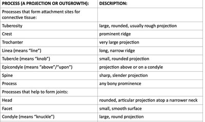
Processes (Projections or Outgrowths): Form attachment sites for connective tissue or help form joints.
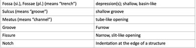
Depressions and Openings: Allow passage of blood vessels and nerves or serve as articulation points.
Table: Bone Markings and Descriptions
Process (Projection or Outgrowth) | Description |
|---|---|
Tuberosity | Large, rounded, usually rough projection |
Crest | Prominent ridge |
Trochanter | Very large projection |
Linea (means "line") | Long, narrow ridge |
Tubercle (means "knob") | Small, rounded projection |
Epicondyle (means "above"/"upon") | Projection above or on a condyle |
Spine | Sharp, slender projection |
Process | Any bony prominence |
Processes that help to form joints | |
Head | Rounded, articular projection atop a narrower neck |
Facet | Small, smooth surface |
Condyle (means "knuckle") | Large, round projection |
Depression or Opening | Description |
|---|---|
Fossa (si.), Fossae (pl.) (means "trench") | Depression(s); shallow, basin-like |
Sulcus (means "groove") | Shallow groove |
Meatus (means "channel") | Tube-like opening |
Groove | Furrow |
Fissure | Narrow, slit-like opening |
Notch | Indentation at the edge of a structure |
Skull Anatomy
Cranial Bones
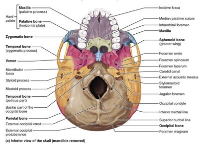
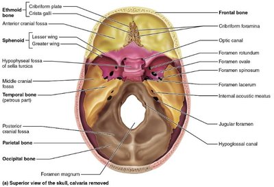
The skull is composed of 8 cranial bones and 14 facial bones, most of which are flat bones. Cranial bones protect the brain and provide attachment sites for head and neck muscles.
Frontal Bone
Parietal Bones
Occipital Bone
Temporal Bones
Sphenoid Bone
Ethmoid Bone
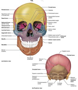
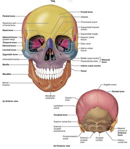
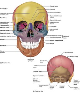
Facial Bones
Facial bones form the structure of the face and house the teeth.
Maxillae (Maxillary Bones)
Zygomatic Bones
Nasal Bones
Lacrimal Bones
Vomer
Inferior Nasal Conchae
Palatine Bones
Mandible
Ethmoid Bone Features
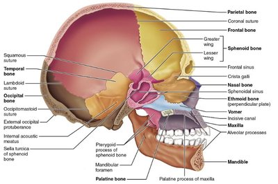
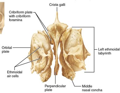
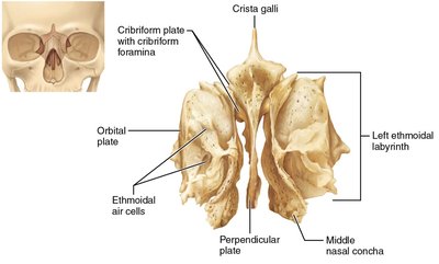
The ethmoid bone is a complex bone located between the eyes, contributing to the medial wall of the orbit, the nasal cavity, and the nasal septum.
Perpendicular Plate: Forms part of the nasal septum.
Middle Nasal Conchae: Increase surface area in the nasal cavity.
Cribriform Plate: Contains foramina for olfactory nerves.
Crista Galli: Attachment for the falx cerebri (dura mater).
Processes and Fossae of the Skull
Various processes and fossae are found throughout the skull, serving as attachment points and passageways.
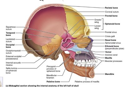
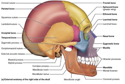
Alveolar Processes: Hold teeth in the maxilla and mandible.
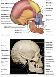
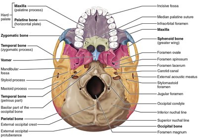
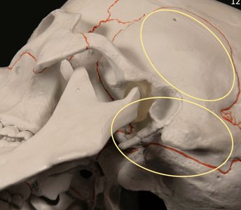
Zygomatic Arches: Formed by the zygomatic bone and the zygomatic process of the temporal bone.
Mastoid and Styloid Processes: Located on the temporal bone, serve as muscle attachment sites.
Mandibular Fossa: Articulates with the condylar process of the mandible.
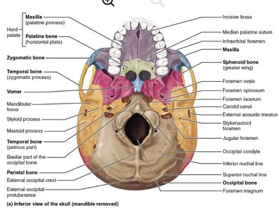
Occipital Condyles: Articulate with the first cervical vertebra.
External Occipital Protuberance: Prominent projection on the occipital bone.
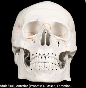
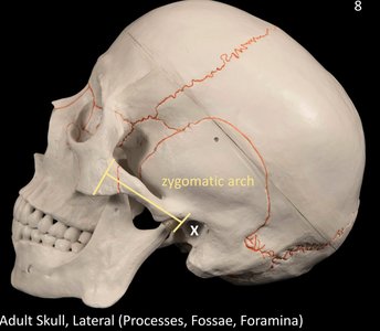
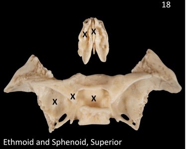
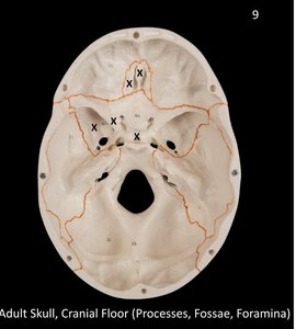
Foramina of the Skull
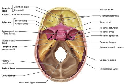
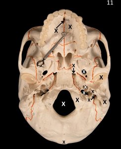
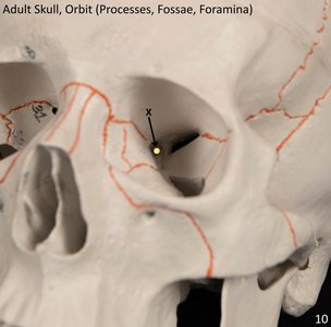
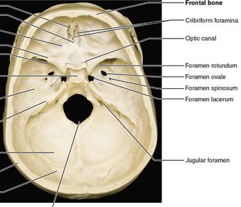
Foramina are openings in bones that allow the passage of nerves and blood vessels.
Foramen Magnum: Large opening in the occipital bone for the spinal cord.
Optic Canal (Optic Foramen): Passage for the optic nerve.
Jugular Foramen: Passage for the jugular vein and cranial nerves.
Carotid Canal: Passage for the internal carotid artery.
Foramen Lacerum, Ovale, Incisive Canal, Stylomastoid Foramen, External Acoustic Meatus: Various passageways for nerves and vessels.
Mandible Anatomy
The mandible is the largest and strongest bone of the face, forming the lower jaw.
Body: Horizontal portion.
Ramus (pl. Rami): Vertical portion.
Angle: Junction of body and ramus.
Coronoid and Condylar Processes: Serve as muscle attachment and articulation points.
Mental Foramina: Openings for nerves and vessels.
Mandibular Foramen: Passage for the inferior alveolar nerve.
Temporomandibular Joint (TMJ)
The TMJ is the articulation between the condylar process of the mandible and the mandibular fossa of the temporal bone, allowing movement of the jaw.
Sutures of the Skull
Sutures are immovable joints that unite the bones of the skull.
Coronal Suture: Between frontal and parietal bones.
Sagittal Suture: Between parietal bones.
Squamous Suture: Between parietal and temporal bones.
Lambdoid Suture: Between parietal and occipital bones.
Fetal and Infant Skull
The fetal and infant skull differs from the adult skull in several ways, allowing for rapid brain growth and flexibility during birth.
Fontanelles: Soft spots where bones are not fully fused.
Flexible Sutures: Allow for expansion.
Large Braincase: Compared to the face.
Fontanelles
Anterior Fontanelle
Sphenoidal (Anterolateral) Fontanelle
Mastoid (Posterolateral) Fontanelle
Posterior Fontanelle
Summary Table: Skull Bones and Markings
Bone | Key Markings/Features |
|---|---|
Frontal | Supraorbital foramen, frontal sinus |
Parietal | Sagittal suture, coronal suture |
Occipital | Foramen magnum, occipital condyles, external occipital protuberance |
Temporal | Mastoid process, styloid process, external acoustic meatus, mandibular fossa |
Sphenoid | Sella turcica, greater/lesser wings, optic canal |
Ethmoid | Cribriform plate, crista galli, perpendicular plate, middle nasal concha |
Maxilla | Alveolar process, infraorbital foramen, palatine process |
Mandible | Body, ramus, angle, coronoid process, condylar process, mental foramen |