 Back
Chapter 3: Dermatology and the Integumentary System – Medical Terminology Study Notes
Study Guide - Smart Notes
 Tailored notes based on your materials, expanded with key definitions, examples, and context.
Dermatology and the Integumentary System
Introduction to Dermatology
Dermatology is the medical specialty focused on the anatomy, physiology, diseases, and treatment of the integumentary system, which includes the skin, hair, nails, and subcutaneous tissue. Dermatologists utilize laboratory tests, diagnostic procedures, medical and surgical interventions, and pharmacological treatments to manage integumentary diseases.

Anatomy of the Integumentary System
Overview of Structures
The integumentary system covers most of the body’s surface and is composed of the skin (the largest organ), nails, hair, and subcutaneous tissue. The term integumentary is derived from integument/o- (skin) and -ary (pertaining to). The term cutaneous also means pertaining to the skin.
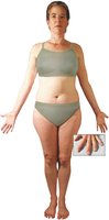
Layers of the Skin
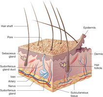
Epidermis: The thin, outermost layer made of epithelial tissue. It consists of the squamous layer (surface, dead keratinized cells) and the basal layer (deep, living, dividing cells). The epidermis contains no blood vessels and relies on the dermis for nutrients. It also contains melanocytes, which produce melanin for UV protection.
Dermis: The thick layer beneath the epidermis, composed of connective tissue, collagen, and elastin. It contains blood vessels, nerves, hair follicles, sebaceous (oil) glands, and sudoriferous (sweat) glands.
Subcutaneous Tissue (Sub Q): Loose connective tissue beneath the dermis, composed mainly of adipose (fat) tissue and lipocytes. It cushions, insulates, and protects internal organs.
Accessory Structures
Hair: Covers most of the body, with color determined by melanocytes. Each hair grows from a follicle in the dermis and is composed of keratinized cells. Piloerection is the process where hair stands up due to cold or emotional stimuli.
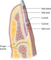
Nails: Consist of the nail plate, root, lunula, cuticle, and nail bed. Nails are formed from living cells in the root that die and harden as they grow outward.
Glands of the Skin
Sebaceous Glands: Exocrine glands that produce sebum (oil), which moisturizes hair and skin.
Sudoriferous Glands: Exocrine glands that secrete sweat, which helps regulate body temperature and excrete waste.
Dermatomes
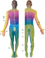
A dermatome is a specific area of skin that sends sensory information to a particular spinal nerve. Each dermatome is named according to the spinal cord level it communicates with (C for cervical, T for thoracic, L for lumbar, S for sacral).
Physiology of the Integumentary System
Protection
The skin acts as the first line of defense against injury and infection.
The acidic surface and keratinized cells discourage microorganism growth and make the skin waterproof.
Sweat and sebum contain antibodies and enzymes that kill bacteria.
Repair
Basal epidermal cells migrate to cover wounds; deeper wounds form clots and scabs, with new cells filling in the area.
Sensation
Sensory receptors in the dermis detect touch, pressure, vibration, pain, and temperature, relaying information to the nervous system.
Vitamin D Synthesis
UV rays convert epidermal cholesterol into vitamin D, which is essential for calcium absorption.
Sun exposure requirements vary by skin tone.
Thermoregulation
Subcutaneous fat conserves heat; piloerector muscles and sweat glands help regulate temperature; dermal blood vessels dilate to release heat.
Homeostasis
The integumentary system maintains internal balance and stability, with skin conditions often signaling systemic issues.
Diseases and Disorders of the Integumentary System
Skin Lesions
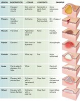
Skin lesions are classified by their appearance, color, contents, and examples. Common types include cysts, fissures, macules, papules, pustules, scales, vesicles, and wheals.
Lesion | Description | Color | Contents | Example |
|---|---|---|---|---|
Cyst | Elevated circular mound | Skin color or erythema | Semisolid or partly fluid-filled | Acne sebaceous cyst |
Fissure | Small, cracklike crevice | Erythema | None/some fluid exudate | Dry, chapped skin |
Macule | Flat circle | Pigmented brown or black | None | Freckle, age spot |
Papule | Elevated | Skin color or erythema | Solid | Acne pimple |
Pustule | Elevated | White top | Pus | Acne whitehead |
Scale | Flat to slightly elevated, thin flake | White | None | Dandruff, psoriasis |
Vesicle | Elevated with pointed top | Erythema with transparent top | Clear fluid | Herpes, chickenpox, shingles |
Wheal | Elevated with broad, flat top | Erythema with pale top | Clear fluid | Insect bites, urticaria |
Skin Color Disorders
Albinism: Normal number of melanocytes but little or no melanin production, resulting in very light skin, hair, and eyes.
Cyanosis: Bluish discoloration due to low oxygen levels, commonly seen in newborns but resolves with oxygenation.
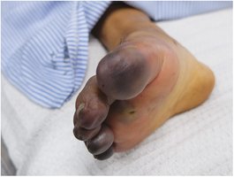
Necrosis: Tissue death due to lack of blood supply, often following severe injury or frostbite.
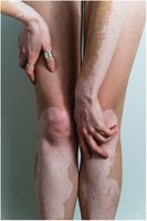
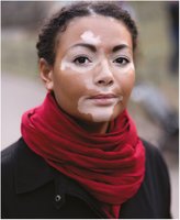
Vitiligo: Autoimmune disorder causing progressive depigmentation in irregular patches, more noticeable in darker skin.


Skin Injuries
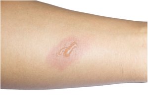
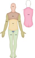
Burns: Classified by depth and extent. Partial-thickness burns cause blisters and fluid-filled bullae. The "Rule of Nines" estimates burn surface area for treatment planning.
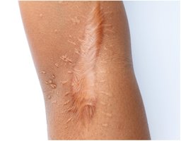
Keloid: Overgrowth of scar tissue beyond the original wound.
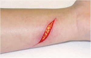
Laceration: Deep cut penetrating the epidermis and dermis, exposing subcutaneous tissue.
Pressure Injury: Localized damage from prolonged pressure, often in immobile patients.
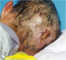
Skin Infections and Infestations
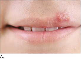
Herpes Simplex Type 1: Causes vesicles (cold sores) on the lips.
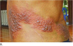
Shingles (Herpes Zoster): Vesicles and crusts along a dermatome, caused by reactivation of the varicella-zoster virus.
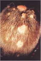
Tinea Capitis (Ringworm): Fungal infection of the scalp, causing round lesions, itching, and hair loss.
Allergic and Inflammatory Skin Conditions
Contact Dermatitis: Inflammatory reaction to allergens or irritants, causing redness, swelling, and sometimes blistering.
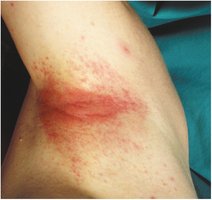
Benign and Malignant Neoplasms
Hemangioma: Benign tumor of dilated blood vessels, appearing as a bright red lesion.
Nevus (Mole): Benign pigmented lesion; can be raised, flat, or resemble a port-wine stain.
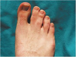
Syndactyly: Congenital fusion of fingers or toes.
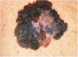
Malignant Melanoma: Cancerous growth of melanocytes, characterized by asymmetry, irregular borders, color variation, and increasing size.
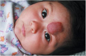
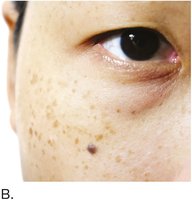
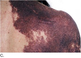
Autoimmune and Chronic Skin Disorders
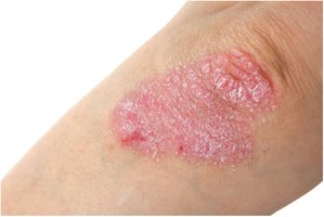
Psoriasis: Chronic autoimmune disorder causing red, scaly plaques, commonly on elbows and knees.
Diseases of the Sebaceous and Sudoriferous Glands
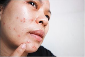
Acne Vulgaris: Inflammation of sebaceous glands, common in adolescence, presenting as papules and pustules.
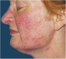
Rosacea: Chronic facial redness and visible blood vessels, often affecting fair-skinned individuals.
Diseases of the Nails
Onychomycosis: Fungal infection of the nails, causing discoloration, thickening, and separation from the nail bed.
Laboratory, Diagnostic, and Medical Procedures
Common Procedures
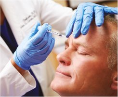
Botox Injection: Use of botulinum toxin to reduce wrinkles by paralyzing facial muscles.
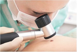
Skin Examination: Visual and magnified inspection of skin lesions, often followed by biopsy for diagnosis.
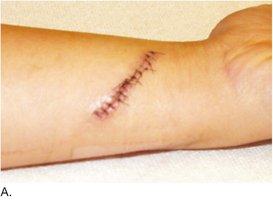
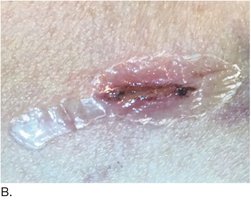
Wound Closure: Lacerations may be closed with sutures or surgical glue, depending on size and location.
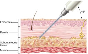
Hypodermic Injection: Administration of drugs into the subcutaneous tissue using a needle at a 45° angle.
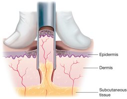
Punch Biopsy: Removal of a cylindrical core of tissue for diagnostic analysis.
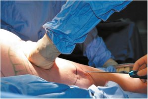
Liposuction: Surgical removal of subcutaneous fat for cosmetic purposes.
Summary Table: Types of Skin Lesions
Lesion | Description | Color | Contents | Example |
|---|---|---|---|---|
Cyst | Elevated circular mound | Skin color or erythema | Semisolid or partly fluid-filled | Acne sebaceous cyst |
Fissure | Small, cracklike crevice | Erythema | None/some fluid exudate | Dry, chapped skin |
Macule | Flat circle | Pigmented brown or black | None | Freckle, age spot |
Papule | Elevated | Skin color or erythema | Solid | Acne pimple |
Pustule | Elevated | White top | Pus | Acne whitehead |
Scale | Flat to slightly elevated, thin flake | White | None | Dandruff, psoriasis |
Vesicle | Elevated with pointed top | Erythema with transparent top | Clear fluid | Herpes, chickenpox, shingles |
Wheal | Elevated with broad, flat top | Erythema with pale top | Clear fluid | Insect bites, urticaria |
Key Medical Terminology
integument/o-: skin
cutane/o-: skin
dermat/o-: skin
-ary: pertaining to
-ous: pertaining to
melan/o-: black
-cyte: cell
pil/o-: hair
erect/o-: stand up
-ion: action or condition
Additional info: This guide expands on the provided lecture slides and textbook images, integrating academic context and terminology for comprehensive exam preparation in Medical Terminology.
