 Back
BackComprehensive Study Notes: Oncology and Cancer Terminology
Study Guide - Smart Notes
 Tailored notes based on your materials, expanded with key definitions, examples, and context.
Oncology: Introduction and Overview
Definition and Scope
Oncology is the medical specialty focused on the study, diagnosis, and treatment of cancer. Unlike other specialties, oncology is not limited to a single body system, as cancer can arise in any tissue or organ. The field encompasses the anatomy and physiology of cancer cells, laboratory and diagnostic tests, medical and surgical procedures, and pharmacological treatments.
Cell Structure and Function
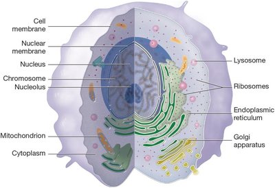
Structures of a Normal Cell
Cells are the smallest independently functioning units in the body, capable of reproduction through division. Each cell contains specialized structures (organelles) that perform essential functions such as nutrient uptake, energy production, protein synthesis, and defense against pathogens.
Cell membrane: Regulates entry and exit of substances.
Nucleus: Contains genetic material (chromosomes).
Nucleolus: Site of ribosome synthesis.
Mitochondrion: Produces cellular energy (ATP).
Endoplasmic reticulum: Synthesizes proteins and lipids.
Golgi apparatus: Modifies and packages proteins.
Lysosome: Digests cellular waste.
Ribosomes: Sites of protein synthesis.
Cytoplasm: Gel-like substance containing organelles.
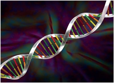
Genetic Material: Chromosomes, DNA, and Genes
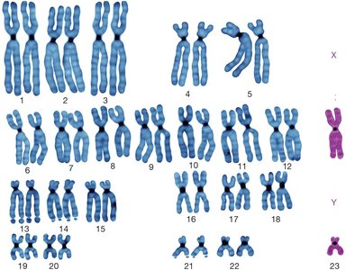
Chromosomes are paired structures within the nucleus, each containing a long DNA molecule. Humans have 46 chromosomes (23 pairs). Genes are segments of DNA that encode instructions for making proteins, which determine cell structure and function.
Cell Division: Normal and Cancerous
Normal Cell Division
Normal cells divide by mitosis in an orderly manner, typically in response to growth signals or tissue repair needs. Suppressor genes in DNA inhibit excessive cell division, maintaining tissue homeostasis. Meiosis is a specialized division producing gametes (sperm and ova) with half the chromosome number (23), ensuring genetic diversity.
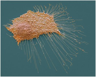
Cancer Cell Division and Growth
Cancer cells arise when normal regulatory mechanisms fail. They may lose differentiation (specialized function), revert to an immature state (anaplasia), and divide uncontrollably. Key terms include:
Differentiation: Process by which cells become specialized.
Undifferentiation: Loss of specialized features; cells appear immature.
Hyperplasia: Increased number of normal cells.
Dysplasia: Abnormal cell growth and arrangement.
Anaplasia: Complete loss of differentiation; cells are primitive.
Causes of Cancer
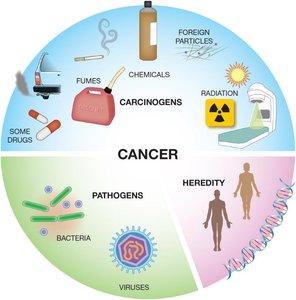
Etiology
Cancer can be caused by a variety of factors, including environmental carcinogens, pathogens, and hereditary mutations. Carcinogens include chemicals, radiation, and certain drugs. Pathogens such as bacteria and viruses can also initiate cancerous changes. Inherited genetic mutations (e.g., in tumor suppressor genes) increase cancer risk.
Incidence and Epidemiology
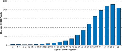
Cancer Diagnosis Across the Lifespan
The risk of developing cancer increases with age. While cancer is rare in children and young adults, incidence rises sharply in older populations. Nearly 40% of people will be diagnosed with cancer at some point in their lives.
Medical Terminology in Oncology
Key Terms and Definitions
Anaplasia: Condition where mature, differentiated cells become undifferentiated and cancerous.
Carcinoid tumor: Slow-growing tumor, often in the digestive tract, rarely metastasizes.
Carcinomatosis: Presence of cancerous tumors at multiple body sites.
Dysplasia: Abnormal cell size, shape, or organization; not yet cancerous.
Lymphadenopathy: Enlarged lymph nodes, often due to trapped cancer cells.
Precancerous: Abnormal cells/tissues not yet fully cancerous.
Characteristics of Cancer Cells and Tumors
Eight Hallmarks of Cancer Cells
Cancer cells differ from normal cells in several key ways:
Do not contribute to normal body function.
Are undifferentiated and lack specialized function.
Are disorganized in arrangement.
Divide more rapidly than normal cells.
Form irregular, unencapsulated solid tumors.
Induce angiogenesis (growth of new blood vessels).
Are invasive, infiltrating surrounding tissues.
Can metastasize (spread) to distant sites via blood or lymph.
Warning Signs and Progression of Cancer
Warning Signs
Change in bowel or bladder habits
Non-healing sores
Unusual bleeding or discharge
Lumps or thickening
Indigestion or swallowing difficulty
Changes in warts or moles
Persistent cough or hoarseness
Unexplained anemia or weight loss
Progression Terms
Neoplasm: Any new, abnormal tissue growth (benign or malignant).
Metastasis: Spread of cancer cells to distant sites.
Remission: Period with no cancer symptoms or signs.
Relapse: Return of cancer after improvement.
Palliative care: Symptom management when cure is not possible.
Classification of Cancers by Body System
Common Cancers and Their Systems
Cancer | Body System |
|---|---|
Bladder cancer | Urinary |
Brain cancer | Nervous |
Breast cancer | Female genital |
Colon cancer | Gastrointestinal |
Leukemia | Blood |
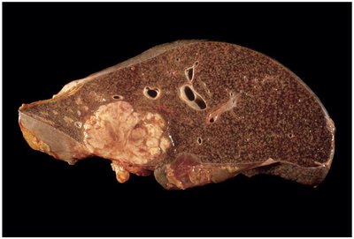
Liver cancer | Gastrointestinal |
Lung cancer | Pulmonary |
Lymphoma | Lymph nodes |
Melanoma | Skin (melanocytes) |
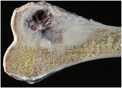
Osteosarcoma | Skeletal |
Ovarian cancer | Female genital |
Renal cell cancer | Urinary |
Retinoblastoma | Eye |
Rhabdomyosarcoma | Muscular |
Stomach cancer | Gastrointestinal |
Testicular cancer | Male genital |
Thyroid cancer | Endocrine |
Wilms tumor | Urinary |
Types of Cancer
Cancer of Blood Cells and Lymphatic Tissue
Leukemia: Cancer of white blood cells (leukocytes).
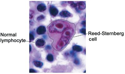
Lymphoma: Cancer of lymph nodes or lymphocytes (e.g., Hodgkin and Non-Hodgkin lymphoma).
Multiple myeloma: Cancer of plasma cells in bone marrow.
Carcinoma
Carcinomas are cancers of epithelial cells, found in skin or mucous membranes. Subtypes include:
Adenocarcinoma: Cancer of glandular epithelial cells (e.g., breast, liver).
Bronchogenic carcinoma: Cancer of bronchial mucous membranes.
Endometrial carcinoma: Cancer of uterine lining.
Small cell carcinoma: Small, round/oval lung cancer cells.
Squamous cell carcinoma: Cancer of flat skin cells, often sun-exposed.
Transitional cell carcinoma: Cancer of urinary tract lining.
Embryonal Cell Cancers (Children)
Hepatoblastoma: Liver cancer in children.
Neuroblastoma: Nerve cell cancer in children.
Retinoblastoma: Retinal cancer in children.
Wilms tumor: Kidney cancer in children.
Sarcoma
Sarcomas are cancers of connective tissues (muscle, bone, cartilage, fat, nerves). They grow rapidly and often metastasize via the bloodstream.
Angiosarcoma: Blood or lymph vessel cancer.
Chondrosarcoma: Cartilage cancer.
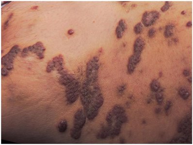
Kaposi sarcoma: Skin/subcutaneous tissue cancer, common in immunocompromised patients.
Leiomyosarcoma: Smooth muscle cancer.
Liposarcoma: Fatty tissue cancer.
Osteosarcoma: Bone cancer.
Rhabdomyosarcoma: Skeletal muscle cancer.
Laboratory and Diagnostic Procedures
Blood Tests
Alpha fetoprotein (AFP): Marker for liver, ovarian, or testicular cancer.
BRCA1/BRCA2: Genetic tests for breast/ovarian cancer risk.
Beta-2 microglobulin: Marker for leukemia, lymphoma, multiple myeloma.
Cancer antigens (CA 15-3, 27-29, 125): Monitor breast/ovarian cancer treatment.
Carcinoembryonic antigen (CEA): Marker for colon, stomach, lung, pancreas cancer.
Complete blood count (CBC): Counts blood cell types.
Human chorionic gonadotropin (HCG): Marker for ovarian/testicular cancer.
Prostate-specific antigen (PSA): Marker for prostate cancer.
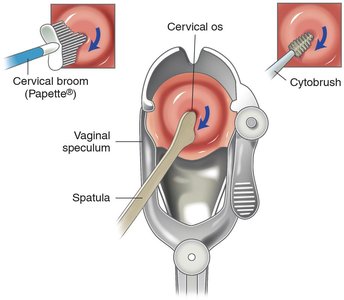
Cytology Tests
Bone marrow aspiration: Diagnoses blood cancers.
Cologuard®: DNA test for colon cancer in stool.
Exfoliative cytology: Examines cells from secretions or tissue scrapings.
Frozen section: Rapid intraoperative tissue diagnosis.
Grading and staging: Classifies cancer by cell appearance and spread.
HER2/neu: Tumor marker test for certain cancers.
Karyotype: Chromosome analysis for abnormalities.
Oncogenomics: Studies cancer-related gene mutations.
Receptor assays: Measures hormone receptor status in breast cancer.
Urine Tests
Urinalysis (UA): Detects cancer markers such as Bence Jones protein, VMA, and 5-HIAA.
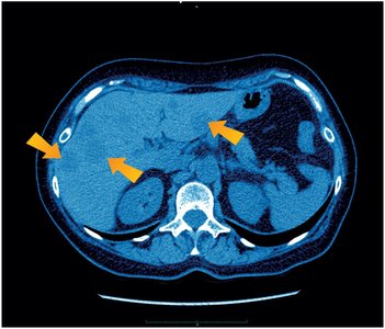
Radiology and Nuclear Medicine Procedures
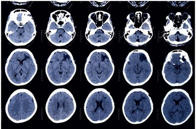
Computerized axial tomography (CAT/CT): X-ray imaging for tumor location and metastasis.
Lymphangiography: X-ray with contrast dye to visualize lymphatic system.
Magnetic resonance imaging (MRI): Uses magnetic fields and radio waves for detailed images.
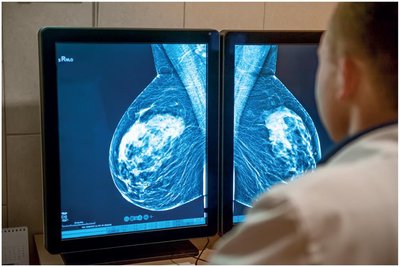
Mammography: X-ray imaging of the breast.
Scintigraphy: Uses radioactive drugs and gamma camera for imaging.
Ultrasonography: Uses sound waves to distinguish cysts from solid tumors.
Cancer Treatments
Chemotherapy
Chemotherapy uses drugs to kill or inhibit cancer cells. Types include:
Alkylating agents: Break DNA strands.
Angiogenesis inhibitors: Block new blood vessel growth.
Antimetabolites: Block DNA synthesis.
Biological response modulators: Stimulate immune response.
Enzyme drugs: Deprive cancer cells of essential amino acids.
Demethylating agents: Inhibit abnormal DNA methylation.
Hormonal drugs: Alter hormonal environment.
Kinase inhibitors: Block cell division enzymes.
Mitosis inhibitors: Disrupt cell division.
Monoclonal antibodies: Target specific cancer cell antigens.
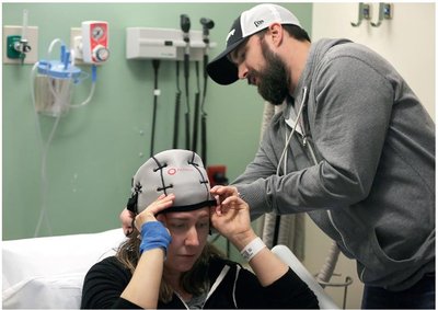
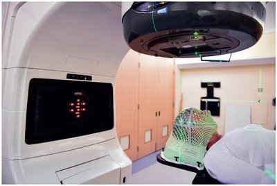
Radiation Therapy
Radiation therapy uses high-energy waves or particles to damage cancer cell DNA. It can be delivered externally (external beam radiotherapy) or internally (brachytherapy, intravenous radiotherapy). Some cancers are radiosensitive, while others are radioresistant.
Medical and Surgical Procedures
Immune cell therapy: Uses patient's own immune cells to attack cancer.
Photodynamic therapy: Uses light-activated drugs to kill cancer cells.
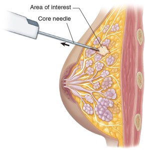
Biopsy (Bx): Removal of tissue for diagnosis (core needle, excisional, fine-needle, incisional, punch, sentinel node).
Bone marrow harvest/transplantation: Replaces destroyed marrow with donor cells.
Cryosurgery: Freezes and destroys small tumors.
Debulking: Removes part of a large tumor.
Electrosurgery: Uses electrical current to destroy tumors.
En bloc resection: Removes tumor and surrounding tissue as a block.
Endoscopy: Visualizes and biopsies internal tissues.
Excision: Removes all or part of a tumor.
Exenteration: Removes tumor and nearby organs.
Exploratory laparotomy: Opens abdomen to explore for cancer.
PICC line insertion: For chemotherapy administration.
Lumpectomy: Removes small breast tumor with margin.
Lymph node dissection: Removes lymph nodes for cancer staging.
Mohs surgery: Removes skin cancer layer by layer.
Percutaneous radiofrequency ablation: Destroys small tumors with heat.
Radical resection: Removes tumor, lymph nodes, and surrounding tissue.
Abbreviations in Oncology
Abbreviation | Definition |
|---|---|
AFP | Alpha fetoprotein |
BRCA | Breast cancer gene |
Bx | Biopsy |
CAT, CT | Computerized axial tomography |
CBC | Complete blood count |
CEA | Carcinoembryonic antigen |
HCG | Human chorionic gonadotropin |
HER2 | Human epidermal growth factor receptor 2 |
MRI | Magnetic resonance imaging |
NED | No evidence of disease |
PICC | Peripherally inserted central catheter |
PR | Progesterone receptor |
PSA | Prostate-specific antigen |
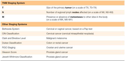
TNM | Tumor, nodes, metastases |
VMA | Vanillylmandelic acid |
