 Back
BackHeart and Neck Vessels: Medical Terminology Study Notes
Study Guide - Smart Notes
 Tailored notes based on your materials, expanded with key definitions, examples, and context.
Heart and Neck Vessels
Precordium and Great Vessels
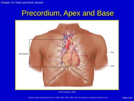
The precordium is the area on the anterior chest directly overlying the heart and great vessels. The heart is located in the mediastinum, between the lungs, and extends from the second to the fifth intercostal space, from the right sternal border to the left midclavicular line. The great vessels include the aorta, pulmonary arteries, and veins, which are responsible for transporting blood to and from the heart.
Layers of the Heart
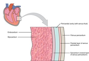
The heart wall consists of three main layers, each with distinct functions:
Pericardium: A double-walled sac that encloses the heart, providing protection and anchorage.
Epicardium: The outermost layer of the heart wall, also known as the visceral layer of the serous pericardium.
Myocardium: The thick, muscular middle layer responsible for the heart's contractile force.
Endocardium: The innermost layer, lining the heart chambers and valves.
Chambers and Valves of the Heart
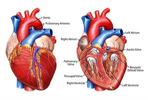
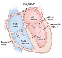
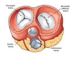
The heart is divided into four chambers and contains four main valves that ensure unidirectional blood flow:
Chambers: Right atrium, right ventricle, left atrium, left ventricle.
Valves:
Atrioventricular (AV) valves: Tricuspid (right), Mitral/Bicuspid (left)
Semilunar (SL) valves: Pulmonic (right), Aortic (left)
Blood Flow Through the Heart
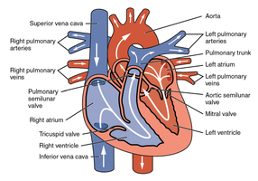
Blood flows through the heart in a specific sequence, ensuring oxygenation and systemic circulation:
Deoxygenated blood enters the right atrium via the superior and inferior vena cava.
Passes through the tricuspid valve into the right ventricle.
Pumped through the pulmonic valve into the pulmonary arteries to the lungs for oxygenation.
Oxygenated blood returns via pulmonary veins to the left atrium.
Passes through the mitral valve into the left ventricle.
Pumped through the aortic valve into the aorta and systemic circulation.
Cardiac Cycle
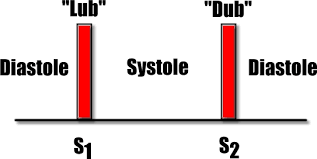
The cardiac cycle describes the rhythmic contraction and relaxation of the heart:
Diastole: Ventricles relax and fill with blood.
Systole: Ventricles contract and pump blood out of the heart.
Heart Sounds
Heart sounds are produced by the closing of heart valves:
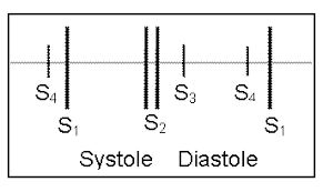
S1 ("Lub"): Closure of AV valves (tricuspid and mitral); marks the beginning of systole.
S2 ("Dub"): Closure of semilunar valves (aortic and pulmonic); marks the end of systole.
S3: Heard just after S2; may be normal in children and young adults, but abnormal in older adults (heart failure).
S4: Heard just before S1; usually abnormal in adults (hypertension).
Heart Murmurs
A murmur is a gentle, blowing, or swooshing sound caused by turbulent blood flow in the heart or great vessels. Murmurs are graded by intensity:
Grade | Description |
|---|---|
1 | Barely audible |
2 | Faint, but audible |
3 | Moderately loud, easy to hear |
4 | Loud, associated with a thrill |
5 | Very loud, audible with stethoscope lifted off chest wall |
6 | Loudest, audible with entire stethoscope lifted off chest wall |
Pericardial Friction Rub
A pericardial friction rub is a high-pitched, scratchy sound caused by inflammation of the pericardium. It is best heard at the apex and left lower sternal border with the diaphragm of the stethoscope while the patient is sitting up and leaning forward.
Electrical Conduction System
The heart's electrical conduction system coordinates contraction:
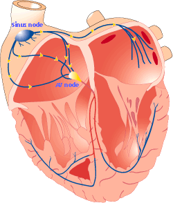
SA node (sinoatrial node): Pacemaker of the heart, initiates impulse.
AV node (atrioventricular node): Delays impulse before passing to ventricles.
Bundle of His, right and left bundle branches, Purkinje fibers: Distribute impulse through ventricles.
Pumping Ability
The heart's pumping ability is measured by:
Cardiac output (CO): Volume of blood pumped by the heart per minute.
Stroke volume (SV): Volume of blood pumped per beat.
Preload: Degree of stretch of the heart muscle before contraction.
Afterload: Resistance the heart must overcome to eject blood.
Contractility: Strength of the heart's contraction.
Formula:
$\text{Cardiac Output (CO)} = \text{Stroke Volume (SV)} \times \text{Heart Rate (HR)}$
The Neck Vessels
Carotid Artery and Jugular Veins
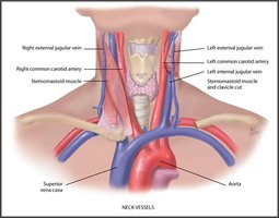
The carotid arteries supply oxygenated blood to the head and neck, while the jugular veins return deoxygenated blood from the head to the heart. Assessment of these vessels provides information about cardiac function and central venous pressure.
Developmental Considerations
Infants and Children
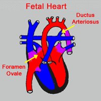
In fetal circulation, the foramen ovale and ductus arteriosus allow blood to bypass the lungs. These structures close shortly after birth as the newborn begins to breathe air.
Pregnant Female
During pregnancy, blood volume and cardiac output increase, while peripheral vasodilation leads to decreased blood pressure.
Aging Adult
Aging affects the cardiovascular system through lifestyle factors and physiological changes, such as increased systolic blood pressure, left ventricular wall thickness, and risk of dysrhythmias.
Subjective Data Collection
Key Questions
Chest pain or tightness (onset, location, duration, characteristics, associated symptoms, relieving factors, timing, severity)
Shortness of breath (dyspnea on exertion, paroxysmal nocturnal dyspnea, orthopnea)
Cough, fatigue, changes in daily activities, cyanosis
Swelling of feet/legs, nocturia
Past medical and family cardiac history
Personal/social history: nutrition, smoking, alcohol, exercise, medications
Objective Data Collection
Carotid Arteries
The carotid arteries are assessed for pulse and the presence of bruits, which may indicate turbulent blood flow due to stenosis or other pathology.
Jugular Venous Pulse
The jugular venous pulse reflects right atrial pressure and can indicate right-sided heart function. Distention may suggest heart failure.
Cardiac Exam
Inspection: Look for precordial pulsations, heaves, or lifts.
Palpation: Assess the apical impulse and precordium for thrills or abnormal movement.
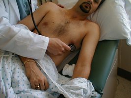
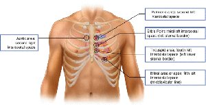
Auscultation: Listen for heart sounds (S1, S2, S3, S4), murmurs, and pericardial friction rubs. Use both the diaphragm and bell of the stethoscope.
Heart Failure: Right vs. Left Sided
Left-Sided Heart Failure | Right-Sided Heart Failure |
|---|---|
|
|
Common Laboratory and Diagnostic Testing
Lipid profile: Assesses cholesterol and triglyceride levels.
Creatinine kinase-MB (CK-MB): Indicates myocardial injury.
Troponin I: Highly specific marker for myocardial infarction.
C-reactive protein: Marker of inflammation.
B-type Natriuretic Peptide (BNP): Elevated in heart failure (normal: 0-99 pg/mL).
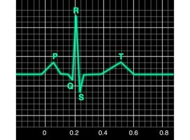
Electrocardiogram (ECG): Records electrical activity of the heart.
Sample Question
Examination of a patient in a supine position reveals distended jugular veins from the base of the neck to the angle of the jaw. This finding indicates which of the following?
Heart Failure
Pneumonia
Hypertension
Cardiovascular Disease
Correct Answer: A. Heart Failure
