Back
BackIntegumentary System: Medical Terminology and Clinical Concepts
Study Guide - Smart Notes
 Tailored notes based on your materials, expanded with key definitions, examples, and context.
Dermatology and the Integumentary System
Introduction to Dermatology
Dermatology is the medical specialty focused on the anatomy, physiology, diseases, and treatment of the integumentary system. This system includes the skin, nails, and subcutaneous tissue, and is essential for protection, sensation, and homeostasis.
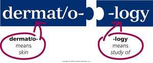
Dermatology: dermat/o- means skin; -logy means study of.
Dermatologists diagnose and treat skin diseases using laboratory tests, medical and surgical procedures, and pharmacological agents.
Anatomy of the Integumentary System
Overview of Structures
The integumentary system covers most of the body and consists of the skin (the largest organ), nails, and subcutaneous tissue.
Skin: Composed of two main layers—epidermis and dermis.
Nails: Protective coverings on the distal ends of fingers and toes.
Subcutaneous tissue: Layer of connective and adipose tissue beneath the dermis.
Word Parts in Medical Terminology
Integument/o-: skin
-ary: pertaining to
Cutane/o-: skin
-ous: pertaining to
Layers of the Skin
Epidermis: Thin, outermost layer made of epithelial tissue; contains dead, keratinized cells at the surface and living, dividing cells at the base (basal layer).
Dermis: Thick layer beneath the epidermis, composed of connective tissue, collagen, and elastin; contains blood vessels, nerves, hair follicles, sebaceous glands, and sweat glands.
Subcutaneous tissue (subQ or subcu): Loose connective and adipose tissue that cushions and insulates the body.
Anatomy of the Epidermis
Squamous layer: Upper part, made of dead cells filled with keratin, providing a protective barrier.
Basal layer: Deep part, made of living cells that divide and move upward; contains melanocytes that produce melanin for UV protection.
Melanocytes: All humans have the same number, but melanin production varies, affecting skin tone and response to sun exposure.
Anatomy of the Dermis
Contains collagen (firm, white protein) and elastin (elastic, yellow protein).
Houses arteries, veins, nerves, hair follicles, sebaceous glands, and sweat glands.
Dermatome: Area of skin sending sensory information to the spinal cord; also refers to a surgical instrument.
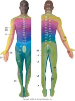
Additional info: Dermatomes are named according to the spinal nerve level (C for cervical, T for thoracic, L for lumbar, S for sacral). The face is innervated by cranial nerves.
Anatomy of the Nails
Nails are composed of both living and dead cells and consist of five main parts: nail plate, nail root, lunula, cuticle, and nail bed.
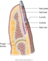
Nail plate: Hard, flat part of the nail.
Nail root: Under the skin, produces keratin-containing cells.
Lunula: White, half-moon at the base of the nail.
Cuticle: Edge around the base of the nail.
Nail bed: Pink tissue under the nail plate.
Anatomy of Hair
Hair covers most of the body; color is determined by melanocytes.
Each hair forms in a follicle in the dermis; hair cells are filled with keratin.
Piloerection: Contraction of a tiny muscle at the hair follicle base causes hair to stand up ("goosebumps").
Anatomy of Sebaceous and Sudoriferous Glands
Sebaceous (oil) glands: Exocrine glands producing sebum, which moisturizes hair and skin.
Sudoriferous (sweat) glands: Exocrine glands secreting sweat (water, sodium, waste) through pores; sweat is odorless until it contacts skin bacteria.
Physiology of the Integumentary System
Protection
The skin acts as the first line of defense against injury and infection.
The acidic environment and keratin discourage microorganism growth and make the skin waterproof.
Sweat and sebum contain antibodies and enzymes that kill bacteria.
Repair
Basal epidermal cells migrate to cover wounds; deeper wounds form clots and scabs.
New cells from the dermis and basal epidermis fill in the wound.
Sensation
Sensory receptors in the dermis detect touch, pressure, vibration, pain, and temperature.
The nervous system relays and interprets these sensations.
Vitamin D Synthesis
UV rays convert epidermal cholesterol into vitamin D, which is essential for calcium absorption.
Thermoregulation
Subcutaneous fat stores heat; piloerector muscles and sweat glands help regulate temperature.
Dermal blood vessels dilate or constrict to release or conserve heat.
Homeostasis
The integumentary system helps maintain internal balance and stability (homeostasis).
Skin conditions can signal other systemic medical issues.
Diseases and Disorders of the Integumentary System
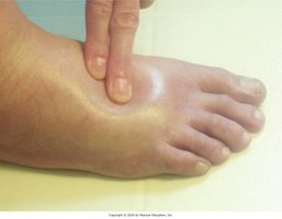
Edema
Edema is swelling caused by excess fluid in the tissues. Pitting edema is identified when pressure leaves a deep indentation.
Types of Skin Lesions
Skin lesions are classified by appearance, color, and contents. Common types include macules, papules, vesicles, pustules, and bullae.
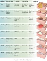
Lesion | Description | Color | Contents | Example |
|---|---|---|---|---|
Vesicle | Small, raised | Erythema | Serous or clear fluid | Herpes, chickenpox |
Pustule | Elevated | White or yellow | Pus | Acne, abscess |
Bulla | Large, raised | Erythema | Serous or clear fluid | Burn, blister |
Macule | Flat, discolored | Pigmented or erythema | None | Freckle, age spot |
Papule | Elevated | Skin color or erythema | None | Wart, mole |
Scale | Flat, thin | White | None | Psoriasis |
Wheal | Elevated, with broad flat top | Erythema | Clear fluid | Allergic reaction |
Necrosis
Necrosis is the death of skin tissue, often due to severe injury, frostbite, or poor blood flow. It may require surgical removal of dead tissue.
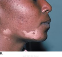
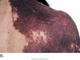
Vitiligo
Vitiligo is a progressive autoimmune disorder causing irregular, expanding areas of depigmentation.
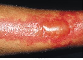
Burns and Skin Injuries
Burns: Damage to the skin caused by heat, chemicals, or electricity. Partial-thickness burns separate the epidermis from the dermis, forming fluid-filled bullae.
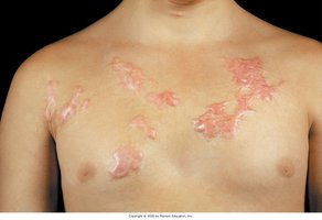
Keloid: An overgrown scar that extends beyond the original injury site.
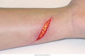
Laceration: A deep cut that penetrates the epidermis and dermis, exposing subcutaneous tissue.
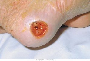
Pressure injury: Localized damage to skin and underlying tissue due to prolonged pressure, often over bony areas.
Skin Infections and Infestations
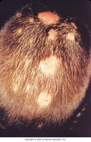
Tinea capitis: Fungal infection (ringworm) of the scalp, causing round lesions, itching, and hair loss.
Allergic Skin Conditions
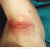
Contact dermatitis: Inflammatory reaction caused by contact with allergens or irritants, resulting in redness and itching.
Benign and Malignant Neoplasms
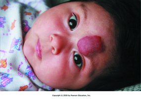
Hemangioma: Benign tumor of dilated blood vessels, appearing as a bright red lesion.
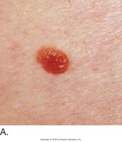
Nevus (mole): Benign pigmented lesion; port-wine stain is a flat, red or purple nevus.
Syndactyly: Congenital fusion of skin and soft tissues between fingers or toes.
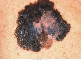
Malignant melanoma: Aggressive skin cancer with asymmetry, irregular borders, color variation, and size > ¼ inch.
Autoimmune Disorders
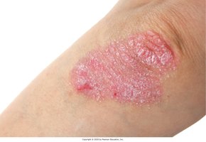
Psoriasis: Chronic autoimmune disease causing erythematous, scaly plaques, commonly on elbows and knees.
Diseases of Sebaceous and Sweat Glands
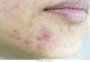
Acne vulgaris: Inflammatory disease of sebaceous glands, common in adolescence.
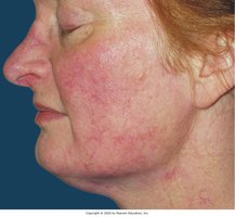
Acne rosacea: Chronic facial redness and dilated blood vessels.
Diseases of the Nails
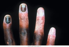
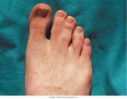
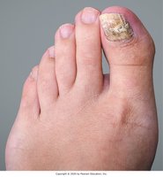
Onychomycosis: Fungal infection of the nails, causing discoloration, thickening, and separation from the nail bed.
Laboratory, Diagnostic, and Medical Procedures
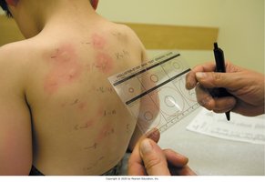
Allergy Skin Testing
Allergy skin testing involves injecting allergens and observing for wheal formation, indicating an immune response.
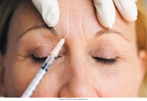
Botox Injection
Botox is a neurotoxin used in small doses to reduce wrinkles by paralyzing facial muscles.
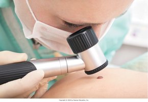
Skin Examination and Biopsy
Dermatologists use magnification to examine lesions and may perform a biopsy for diagnosis.
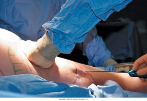
Wound Closure
Lacerations are closed with sutures in layers; deep sutures are absorbable, while skin sutures are removed after healing.

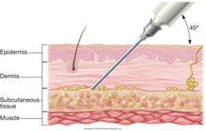
Hypodermic Injection
Medications can be administered into the subcutaneous tissue using a hypodermic needle at a 45-degree angle.
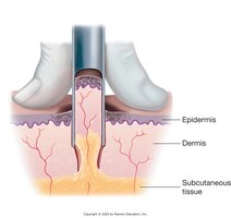
Punch Biopsy
A punch biopsy removes a core of skin tissue, including epidermis, dermis, and subcutaneous tissue, for diagnostic analysis.
Liposuction
Liposuction is a surgical procedure to remove subcutaneous fat for cosmetic purposes.