 Back
BackMedical Terminology: Gastroenterology and the Gastrointestinal System
Study Guide - Smart Notes
 Tailored notes based on your materials, expanded with key definitions, examples, and context.
Gastroenterology: Introduction and Medical Terminology
Definition and Scope
Gastroenterology is the medical specialty focused on the anatomy, physiology, diseases, and treatment of the gastrointestinal (GI) system. Gastroenterologists use diagnostic tests, medical and surgical procedures, and pharmacological therapies to manage GI diseases.
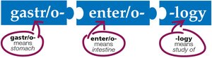
Gastr/o-: Refers to the stomach
Enter/o-: Refers to the intestine
-logy: Means the study of
Anatomy of the Gastrointestinal System
Overview of the GI System
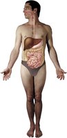
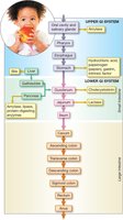
The GI system consists of a series of organs and glands forming a continuous pathway from the mouth to the anus. Its primary functions are digestion, absorption of nutrients, and elimination of waste.
Upper Gastrointestinal System
Oral Cavity and Pharynx
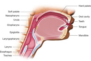
The oral cavity is the entry point of the GI tract and contains the teeth, gums, tongue, hard palate, and soft palate. The pharynx (throat) serves as a common passageway for food and air, divided into the nasopharynx, oropharynx, and laryngopharynx.
Tongue: Contains taste receptors that send signals to the gustatory cortex.
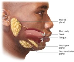
Salivary glands: Three pairs (parotid, sublingual, submandibular) secrete saliva to begin digestion.
Hard palate: Forms the anterior roof of the mouth.
Soft palate and uvula: Form the posterior roof, aiding in swallowing and preventing food from entering the nasal cavity.
Esophagus
The esophagus is a muscular tube connecting the pharynx to the stomach. Peristalsis (coordinated muscular contractions) moves food toward the stomach.
Stomach
The stomach is an elongated sac with four regions: cardia, fundus, body, and pylorus. It mixes food with gastric juices to form chyme. Two sphincters (lower esophageal and pyloric) regulate entry and exit of food.
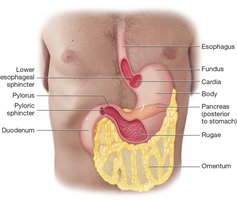
Rugae: Folds in the gastric mucosa that allow expansion.
Chyme: Mixture of food, saliva, and digestive enzymes.
Lower Gastrointestinal System
Small Intestine
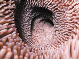
The small intestine is a long, narrow tube divided into three segments: duodenum, jejunum, and ileum. It is lined with villi to maximize nutrient absorption.
Duodenum: First segment, receives chyme from the stomach.
Jejunum: Middle segment, primary site for nutrient absorption.
Ileum: Final segment, absorbs remaining nutrients and water.
Large Intestine
The large intestine absorbs water and forms feces. It consists of the cecum (with appendix), colon (ascending, transverse, descending, sigmoid), rectum, and anus.
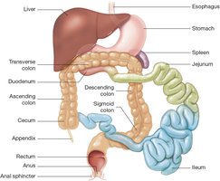
Haustra: Pouches in the colon that expand to accommodate waste.
Ileocecal valve: Prevents backflow from the large intestine to the small intestine.
Anal sphincter: Voluntary muscle controlling defecation.
Abdominopelvic Cavity and Peritoneum
The intestines are housed in the abdominopelvic cavity, lined by the peritoneum, which secretes fluid and forms the omentum and mesentery for organ support.
Accessory Organs
Liver
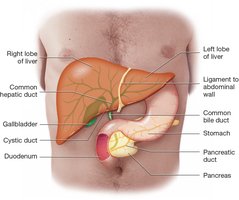
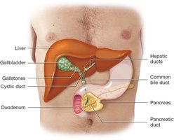
The liver is the largest solid organ, producing bile for fat digestion. Bile flows through the biliary tree into the gallbladder and duodenum.
Gallbladder
The gallbladder stores and concentrates bile, releasing it into the duodenum in response to fatty chyme.
Pancreas
The pancreas secretes digestive enzymes (amylase, lipase, proteases) into the duodenum and also produces insulin as part of the endocrine system.
Physiology of Digestion
Mechanical Digestion
Mechanical digestion involves physical processes such as mastication (chewing), deglutition (swallowing), and peristalsis (muscular movement of food).
Chemical Digestion
Chemical digestion uses enzymes and acids to break down food:
Salivary amylase: Begins carbohydrate digestion in the mouth.
Stomach secretions: Hydrochloric acid, pepsinogen (converted to pepsin for protein digestion), gastrin (stimulates secretions), and intrinsic factor (aids vitamin B12 absorption).
Liver and gallbladder: Bile emulsifies fats.
Pancreas: Amylase (carbohydrates), lipase (fats), proteases (proteins).
Jejunum: Lactase breaks down lactose.
Absorption
Absorption is the process by which nutrients and fluids pass into the bloodstream. Most absorption occurs in the ileum, with water reabsorbed in the large intestine.
Elimination
Elimination removes undigested waste as feces through the process of defecation. Flatus refers to gas expelled from the digestive tract.
Common Diseases of the Gastrointestinal System
Esophagus and Stomach Disorders
Dyspepsia: Indigestion caused by excess acid, reflux, overeating, or stress.

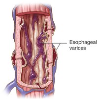
Esophageal varices: Swollen veins in the esophagus that can bleed.
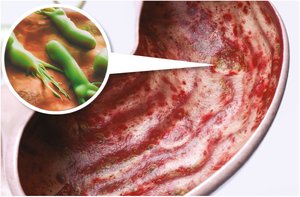
Gastric ulcer: Erosion of the stomach lining, often with bleeding.
Small and Large Intestine Disorders
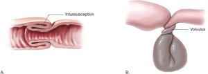
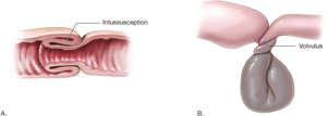
Intussusception: The intestine folds into itself, causing obstruction.
Volvulus: Twisting of the intestine, leading to obstruction and reduced blood flow.
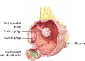
Diverticula: Pouches in the colon wall where feces can become trapped.
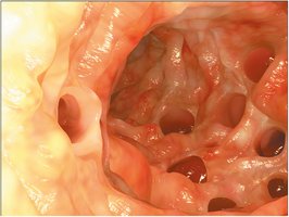
Diverticulitis: Inflammation of a diverticulum due to trapped feces.
Polyposis: Presence of multiple polyps, which can become cancerous.
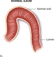
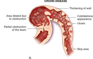
Crohn disease: Chronic inflammation causing thickening and ulceration of the intestinal wall.
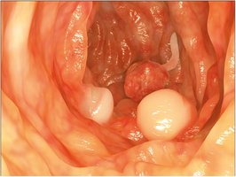
Colonic polyps: Growths in the colon that may be sessile (flat) or pedunculated (on a stalk).
Abdominal Wall and Cavity Disorders
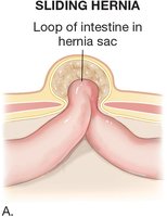
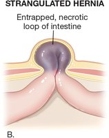
Hernia: Protrusion of intestine through the abdominal wall. Types include sliding and strangulated hernias.
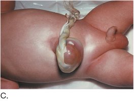
Omphalocele: Congenital hernia at the umbilicus.
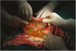
Peritonitis: Inflammation of the peritoneum, often due to perforation and leakage of intestinal contents.
Liver Disorders
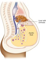
Ascites: Accumulation of fluid in the abdominopelvic cavity due to liver disease.
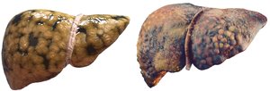
Fatty liver and cirrhosis: Fatty infiltration and scarring of the liver, reducing function.
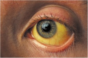
Jaundice: Yellow discoloration of the skin and sclera due to bilirubin buildup.
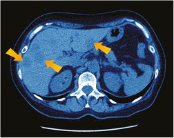
Liver cancer: Malignant tumors in the liver, often detected by imaging.
Gallbladder and Bile Duct Disorders
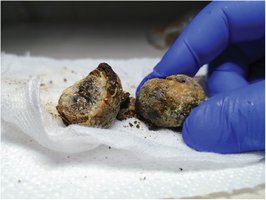
Gallstones (cholelithiasis): Stones that block bile ducts, causing pain and possible infection.
Diagnostic and Medical Procedures
Radiologic and Endoscopic Procedures
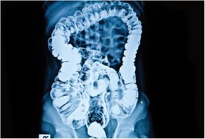
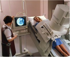
Barium enema: X-ray imaging of the colon after barium contrast is introduced rectally.
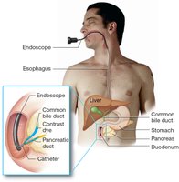
Endoscopic retrograde cholangiopancreatography (ERCP): Endoscopic imaging of the bile and pancreatic ducts using contrast dye.
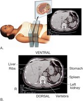
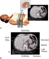
CT scan: Cross-sectional imaging to visualize abdominal organs and detect pathology.
Upper GI series: X-ray imaging after swallowing barium to visualize the upper GI tract.
Medical and Surgical Procedures
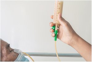
Nasogastric (NG) tube: Inserted through the nose into the stomach for feeding or medication administration.
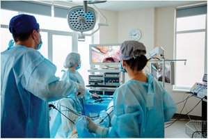
Laparoscopic cholecystectomy: Minimally invasive removal of the gallbladder using a laparoscope.
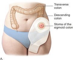
Colostomy and stoma: Surgical creation of an opening from the colon to the abdominal wall for fecal diversion.
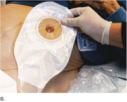
Colonoscopy: Endoscopic examination of the colon and rectum.
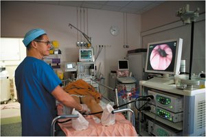
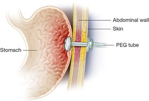
Percutaneous endoscopic gastrostomy (PEG) tube: Permanent feeding tube inserted into the stomach through the abdominal wall.
Additional info: This guide covers the essential medical terminology, anatomy, physiology, common diseases, and diagnostic/surgical procedures relevant to the gastrointestinal system for medical terminology students.
