 Back
BackMuscular System: Structure, Function, and Key Muscles
Study Guide - Smart Notes
 Tailored notes based on your materials, expanded with key definitions, examples, and context.
Muscular System Overview
Introduction to Muscle Anatomy
The muscular system is essential for movement, stability, and posture in the human body. Muscles are attached to bones and work together to produce motion at joints. Understanding muscle terminology and structure is foundational for medical terminology students.
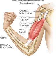
Muscle Belly: The central, fleshy part of a muscle containing skeletal muscle tissue.
Origin: The less movable attachment point of a muscle, typically proximal or closer to the trunk.
Insertion: The more movable attachment point, usually distal; pulled toward the origin during contraction.
Action: The specific movement produced by muscle contraction, such as flexion or extension at a joint.
Agonist and Antagonist Muscles
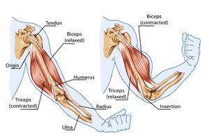
Muscles often work in pairs to control movement. The agonist is the primary muscle responsible for a movement, while the antagonist opposes it.
Agonist: Muscle that causes a specific joint movement (e.g., biceps brachii for elbow flexion).
Antagonist: Muscle that produces the opposite movement (e.g., triceps brachii for elbow extension).
Example: During elbow flexion, the biceps contracts (agonist) and the triceps relaxes (antagonist); during extension, the roles reverse.
Major Muscles of the Human Body
Anterior and Posterior Views
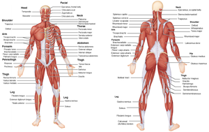
The human body contains numerous muscles, each with specific functions and anatomical locations. Understanding their names and positions is crucial for medical terminology.
Pectoralis major
Deltoid
Trapezius
Serratus anterior
Latissimus dorsi
Biceps brachii
Triceps brachii
Brachioradialis
External oblique
Rectus abdominis
Quadriceps femoris (includes rectus femoris, vastus medialis, vastus lateralis, vastus intermedius)
Tensor fascia latae
Adductor group (adductor longus, adductor magnus, gracilis, etc.)
Hamstrings (semitendinosus, semimembranosus, biceps femoris)
Gluteus maximus
Tibialis anterior
Gastrocnemius
Soleus
Fibularis longus (Peroneus longus)
Muscles of the Shoulder and Upper Limb
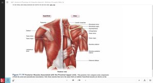
Muscles Moving the Scapula and Humerus (Anterior)
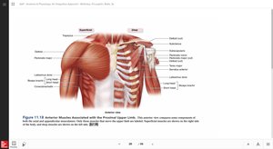
These muscles are responsible for movements of the shoulder and upper arm, including flexion, extension, abduction, and rotation.
Pectoralis major: Adducts and flexes the humerus.
Deltoid: Abducts the arm.
Serratus anterior: Protracts the scapula.
Biceps brachii: Flexes the elbow and supinates the forearm.
Muscles Moving the Scapula and Humerus (Posterior)
Posterior muscles stabilize and move the scapula and humerus, contributing to shoulder extension and rotation.
Trapezius: Elevates, retracts, and rotates the scapula.
Latissimus dorsi: Extends, adducts, and medially rotates the humerus.
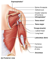
Infraspinatus, teres minor: Lateral rotation of the humerus.
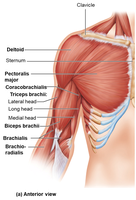
Muscles of the Shoulder and Elbow (Anterior and Posterior Views)
Biceps brachii: Flexes the elbow.
Brachialis: Flexes the elbow.
Brachioradialis: Flexes the forearm.
Triceps brachii: Extends the elbow.
Muscles of the Abdominal Wall
Structure and Function
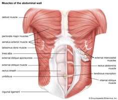
The abdominal wall muscles support the trunk, allow movement, and help maintain posture. They also protect internal organs and assist in breathing.
Rectus abdominis: Flexes the vertebral column.
External oblique: Compresses the abdomen and rotates the trunk.
Internal oblique: Compresses the abdomen and rotates the trunk.
Transversus abdominis: Compresses abdominal contents.
Muscles of the Hip and Knee
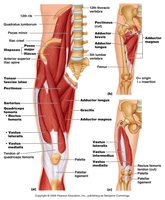
Anterior Muscles
Muscles in the anterior thigh are primarily responsible for hip flexion and knee extension.
Quadriceps femoris: Extends the knee.
Adductor group: Adducts the thigh.
Tensor fascia latae: Abducts and medially rotates the thigh.
Posterior Muscles
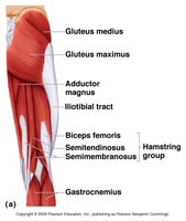
Posterior thigh muscles are involved in hip extension and knee flexion.
Gluteus maximus: Extends and laterally rotates the hip.
Hamstrings: Flex the knee and extend the hip.
Adductor magnus: Adducts and extends the thigh.
Muscles of the Lower Leg
Anterior Muscles
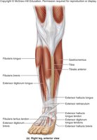
The anterior muscles of the lower leg are responsible for dorsiflexion and extension of the toes.
Tibialis anterior: Dorsiflexes and inverts the foot.
Fibularis longus: Everts the foot.
Extensor digitorum longus: Extends the toes.
Posterior Muscles
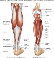
Posterior muscles of the lower leg are involved in plantar flexion and flexion of the toes.
Gastrocnemius: Plantar flexes the foot and flexes the knee.
Soleus: Plantar flexes the foot.
Flexor digitorum longus: Flexes the toes.
Summary Table: Key Muscles and Their Actions
Muscle | Location | Action |
|---|---|---|
Biceps brachii | Upper arm | Elbow flexion |
Triceps brachii | Upper arm | Elbow extension |
Quadriceps femoris | Thigh (anterior) | Knee extension |
Hamstrings | Thigh (posterior) | Knee flexion, hip extension |
Gluteus maximus | Buttocks | Hip extension |
Tibialis anterior | Lower leg (anterior) | Dorsiflexion |
Gastrocnemius | Lower leg (posterior) | Plantar flexion |
Key Medical Terminology
Flexion: Decreasing the angle between two bones (e.g., bending the elbow).
Extension: Increasing the angle between two bones (e.g., straightening the knee).
Abduction: Moving a limb away from the midline.
Adduction: Moving a limb toward the midline.
Rotation: Moving a bone around its longitudinal axis.
Conclusion
Understanding the structure, function, and terminology of muscles is essential for medical terminology students. Mastery of these concepts supports further study in anatomy, physiology, and clinical practice. Additional info: Expanded explanations and context were added to ensure completeness and academic quality.
