 Back
BackMusculoskeletal System: Medical Terminology and Anatomy Study Guide
Study Guide - Smart Notes
 Tailored notes based on your materials, expanded with key definitions, examples, and context.
Musculoskeletal System Overview
Introduction
The musculoskeletal system is a complex network of bones, joints, and muscles that provides structure, protection, movement, and support to the human body. Understanding the terminology, anatomy, and physiology of this system is essential for students in medical and allied health fields.
Skeletal System
General Structure and Function
Bones: The adult human skeleton consists of 206 bones, forming the internal framework of the body.
Functions: Support, protection of organs, muscle attachment, blood cell production (hematopoiesis), and mineral storage (e.g., calcium, phosphorus).
Organs: Bones and joints are considered the organs of the skeletal system.
Bone Structure and Classification
Osseous tissue: The specialized connective tissue that forms bones, among the hardest materials in the body.
Ossification: The process by which cartilage is gradually replaced by bone tissue, beginning in the fetal skeleton and continuing through adolescence.
Remodeling: Continuous process where old bone is broken down by osteoclasts and new bone is formed by osteoblasts; in adults, the skeleton is replaced approximately every 10 years.
Classification by Shape
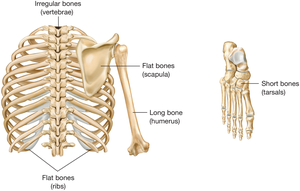
Long bones: e.g., femur, humerus
Short bones: e.g., carpals, tarsals
Flat bones: e.g., scapula, ribs
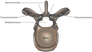
Irregular bones: e.g., vertebrae
Long Bone Anatomy
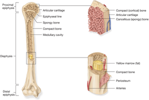
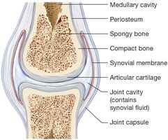
Diaphysis: The central shaft containing the medullary cavity (yellow marrow).
Epiphysis: The wide ends of the bone, covered with articular cartilage for joint movement.
Periosteum: Tough, fibrous membrane covering the bone except at joint surfaces.
Compact (cortical) bone: Dense outer layer providing strength.
Cancellous (spongy) bone: Porous inner bone containing red marrow for blood cell production.
Bony Projections and Depressions
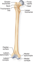
Processes: Projections or outgrowths on bones that serve as attachment points for muscles or form joints.
Examples: Trochanter, tubercle, condyle, crest, spine (projections); fossa, foramen, sinus (depressions).
Divisions of the Skeleton
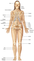
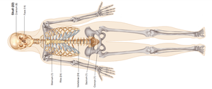
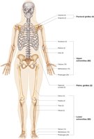
Axial skeleton: Skull, vertebral column, rib cage, and sternum (central axis of the body).
Appendicular skeleton: Pectoral girdle, upper limbs, pelvic girdle, and lower limbs (appendages).
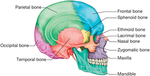
Skull and Facial Bones
Cranium: Frontal, parietal, temporal, ethmoid, sphenoid, occipital bones.
Facial bones: Mandible, maxilla, zygomatic, vomer, palatine, nasal, lacrimal bones.
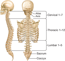
Vertebral Column
Cervical (C1–C7): Neck region
Thoracic (T1–T12): Chest region
Lumbar (L1–L5): Lower back
Sacrum: Base of the spine
Coccyx: Tailbone
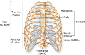
Rib Cage
True ribs (1–7): Attach directly to the sternum
False ribs (8–10): Attach indirectly to the sternum
Floating ribs (11–12): Do not attach to the sternum
Appendicular Skeleton
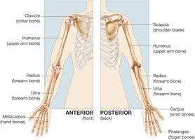
Pectoral girdle: Clavicle and scapula
Upper extremity: Humerus, radius, ulna, carpals, metacarpals, phalanges
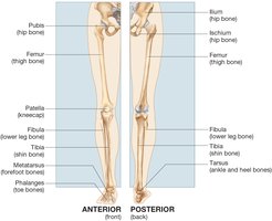
Pelvic girdle: Ilium, ischium, pubis
Lower extremity: Femur, patella, tibia, fibula, tarsals, metatarsals, phalanges
Joints (Articulations)
Definition: Joints are locations where two or more bones meet.
Types of joints:
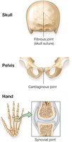
Synovial joints: Freely movable (e.g., knee, shoulder); contain synovial fluid and are stabilized by ligaments and sometimes menisci or bursae.
Cartilaginous joints: Slightly movable, bones joined by cartilage (e.g., pubic symphysis).
Fibrous joints: Immovable, bones joined by fibrous tissue (e.g., skull sutures).
Bone Pathology and Fractures
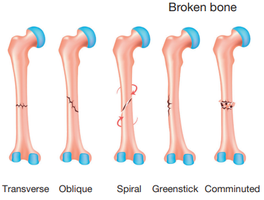
Common Fracture Types
Transverse: Straight across the bone
Oblique: Angled fracture
Spiral: Twisting force causes a spiral break
Greenstick: Incomplete break, common in children
Comminuted: Bone is shattered into several pieces
Bone Diseases
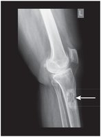
Osteogenic sarcoma: Malignant bone tumor, often seen in long bones.
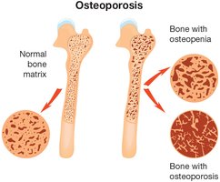
Osteopenia and osteoporosis: Conditions of decreased bone density, increasing fracture risk.
Spinal Pathology
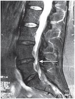
Herniated disk: Protrusion of intervertebral disk, may compress spinal nerves.
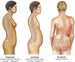
Abnormal curvatures: Kyphosis (humpback), lordosis (swayback), scoliosis (lateral curvature).
Muscular System
General Structure and Function
Muscles: Organs composed of bundles of muscle fibers that contract to produce movement.
Functions: Movement, posture, heat production, and stabilization of joints.
Types of muscle tissue: Skeletal (voluntary), smooth (involuntary), cardiac (involuntary).
Skeletal Muscle Anatomy and Actions
Origin: Less movable attachment point of a muscle.
Insertion: More movable attachment point.
Action: The movement produced by muscle contraction.
Antagonistic pairs: Muscles often work in pairs with opposite actions (e.g., biceps and triceps).
Key Medical Terminology
Combining Forms, Prefixes, and Suffixes
Oste/o: Bone
Arthr/o: Joint
My/o, myos/o: Muscle
-algia: Pain
-itis: Inflammation
-centesis: Surgical puncture to remove fluid
-ectomy: Surgical removal
Hyper-: Excessive
Poly-: Many
Selected Pathology Terms
Osteoarthritis: Inflammation of bones and joints
Kyphosis: Abnormal posterior curvature of the thoracic spine
Myorrhexis: Rupture of muscle
Polymyositis: Inflammation of many muscles
Selected Diagnostic and Therapeutic Terms
Myelography: X-ray imaging of the spinal cord
Arthroscopy: Visual examination of a joint using an endoscope
Arthrocentesis: Puncture of a joint to remove fluid
Bursectomy: Surgical removal of a bursa
Summary Table: Types of Joints
Type of Joint | Movement | Example |
|---|---|---|
Synovial | Freely movable | Knee, shoulder |
Cartilaginous | Slightly movable | Pubic symphysis |
Fibrous | Immovable | Skull sutures |
Conclusion
Mastery of musculoskeletal system terminology, anatomy, and pathology is foundational for medical terminology students. Understanding the structure and function of bones, joints, and muscles, as well as the language used to describe their conditions and treatments, is essential for effective communication in healthcare settings.
