 Back
Neurology: Nervous System Medical Terminology Study Guide
Study Guide - Smart Notes
 Tailored notes based on your materials, expanded with key definitions, examples, and context.
Neurology and the Nervous System
Introduction to Neurology
Neurology is the medical specialty focused on the study, diagnosis, and treatment of the nervous system. It encompasses laboratory and diagnostic tests, medical and surgical procedures, and pharmacological interventions for nervous system diseases.
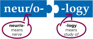
Neurology: The study of nerves and the nervous system.
Practitioners: Neurologists use various tests and procedures to treat nervous system disorders.
Key Medical Terminology:
neur/o-: means nerve
-logy: means study of
Anatomy of the Nervous System
Overview of Nervous System Structure
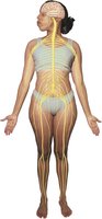
The nervous system is a complex, widespread body system consisting of the brain, spinal cord, and cranial and spinal nerves. It serves as the pathway for nerve impulses throughout the body.
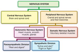
Central Nervous System (CNS): Brain and spinal cord
Peripheral Nervous System (PNS): Cranial and spinal nerves and their branches
Autonomic Nervous System: Controls involuntary functions (heart muscle, smooth muscle, glands)
Somatic Nervous System: Controls voluntary skeletal muscle movements
Divisions:
Parasympathetic Division: "Rest and digest"
Sympathetic Division: "Active and alert" or "Fight or flight"
Anatomy of the Brain
Major Structures of the Brain
The brain is the largest part of the CNS, located in the cranium and composed of several key structures:
Cerebrum: Largest structure, divided into four lobes (frontal, parietal, temporal, occipital) and two hemispheres
Ventricles: Four connected cavities filled with cerebrospinal fluid (CSF)
Thalamus: Relay station for sensory information
Hypothalamus: Regulates temperature, hunger, thirst, emotions
Hippocampus: Transfers short-term memory to long-term memory
Amygdaloid bodies: Interprets facial expressions, links visual images to memory
Brainstem: Connects brain and spinal cord (midbrain, pons, medulla oblongata)
Cerebellum: Maintains balance and coordinates movements
Meninges: Three membrane layers surrounding the brain
Lobes of the Cerebrum
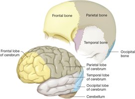
Each lobe of the cerebrum is named after the cranial bone above it and has specialized functions:
Frontal Lobe: Conscious thought, intelligence, prediction, motor control, speech, taste analysis
Parietal Lobe: Touch, temperature, vibration, pain analysis (somatosensory area)
Temporal Lobe: Hearing (auditory cortex), smell (olfactory cortex)
Occipital Lobe: Vision analysis (visual cortex), 3D vision
Brain Hemispheres and Functions
The cerebrum is divided into right and left hemispheres, connected by the corpus callosum. Each hemisphere processes information from the opposite side of the body.
Left Hemisphere: Memories, language, reasoning, problem solving
Right Hemisphere: Environmental analysis, emotion, recognition, artistic abilities

Gyri, Sulci, and Ventricles
The surface of the cerebrum contains folds (gyri) and grooves (sulci). The gray matter forms the outer layer, while white matter lies beneath.
Ventricles: Lined with ependymal cells, produce and circulate CSF
CSF: Cushions and protects the brain
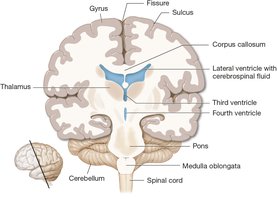
Midline Structures of the Brain
Key midline structures include the thalamus, hypothalamus, midbrain, pons, medulla oblongata, and cerebellum.
Thalamus: Sensory relay station
Hypothalamus: Regulates autonomic functions and emotions
Midbrain: Maintains consciousness, coordinates reflexes
Pons: Relays impulses
Medulla Oblongata: Regulates respiration and heart rate
Cerebellum: Balance and movement coordination
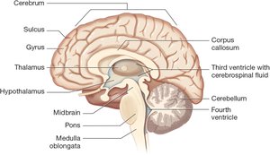
Meninges
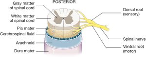
The meninges are three protective membrane layers around the brain:
Dura Mater: Tough, fibrous outer layer
Arachnoid: Cushioning middle layer, surrounds CSF-filled subarachnoid space
Pia Mater: Thin, delicate inner layer with blood vessels
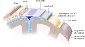
Anatomy of the Spinal Cord
Structure and Protection
The spinal cord is a long, narrow column of tissue beginning at the medulla oblongata and traveling through the spinal canal. It is protected by vertebrae, epidural space, and meningeal layers.
Central Canal: Lined with ependymal cells, produces CSF
Gray Matter: Cell bodies
White Matter: Axons bundled into tracts
Ascending Tracts: Sensory information to the brain
Descending Tracts: Motor commands from the brain
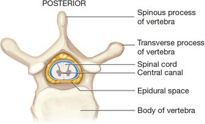
Anatomy of the Cranial and Spinal Nerves
Cranial Nerves
There are twelve pairs of cranial nerves, each with specific sensory and motor functions.
Olfactory: Smell
Optic: Vision
Oculomotor, Trochlear, Abducens: Eye movement
Trigeminal: Facial sensation, chewing
Facial: Taste, tear and saliva secretion
Vestibulocochlear: Hearing, balance
Glossopharyngeal: Taste, blood pressure, swallowing
Vagus: Taste, heart, bronchial, GI muscle control
Accessory: Swallowing, speech, neck movement
Hypoglossal: Tongue movement
Spinal Nerves
There are thirty-one pairs of spinal nerves, named for their adjacent vertebrae. Each nerve has dorsal (sensory) and ventral (motor) roots.
Dorsal Roots: Receive sensory information (afferent)
Ventral Roots: Send motor commands (efferent)
Reflex: Rapid, involuntary muscle reaction controlled by the spinal cord
Divisions of the Nervous System
Autonomic Nervous System
Controls involuntary movements of cardiac and smooth muscle. Divided into:
Parasympathetic Division: Active during rest, uses acetylcholine, decreases heart rate, increases digestion
Sympathetic Division: Active during stress, uses norepinephrine, increases alertness, heart rate, and metabolism
Somatic Nervous System
Controls voluntary movements of skeletal muscles, driven by conscious thought.
Anatomy and Physiology of Neurons and Neuroglia
Neurons
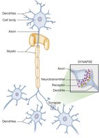
Neurons are individual cells that conduct electrical impulses, forming the functional unit of the nervous system.
Dendrites: Receive impulses
Cell Body: Contains nucleus, produces neurotransmitters
Axon: Transmits impulses, may be covered in myelin
Neuroglia
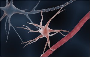
Neuroglia are supportive cells that do not conduct impulses. Five types:
Astrocytes: Form blood-brain barrier
Ependymal Cells: Produce CSF
Microglia: Consume dead tissue and pathogens
Oligodendroglia: Produce myelin in CNS
Schwann Cells: Produce myelin in PNS
Neurotransmitters
Neurotransmitters are chemicals that transmit impulses across synapses.
Acetylcholine: Parasympathetic division
Dopamine: Brain
Endorphins: Brain neuromodulator
Epinephrine: Adrenal hormone
Norepinephrine: Sympathetic division
Serotonin: Brain and spinal cord
Common Nervous System Diseases and Disorders
Brain Tumors
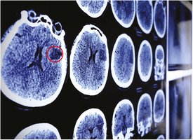
Abnormal growths in the brain, can be benign or malignant. Diagnosis often involves imaging and biopsy.
Astrocytoma
A cancerous tumor arising from astrocytes, a type of neuroglia.
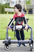
Cerebral Palsy
A disorder affecting movement and muscle tone, often seen in children.
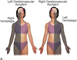
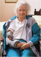
Cerebrovascular Accident (Stroke)
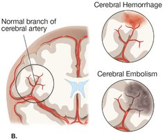
Occurs when blood flow to the brain is disrupted, causing paralysis on the opposite side of the body.
Hemorrhagic: Due to bleeding
Embolic: Due to blockage
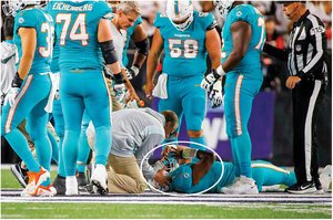
Concussion
A mild traumatic brain injury, often resulting in temporary loss of function.
Down Syndrome
A genetic disorder characterized by distinctive physical features and intellectual disability.

Dyslexia
A learning disorder affecting reading and writing abilities.

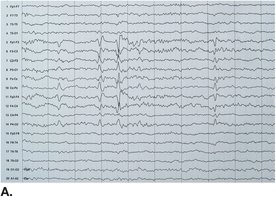
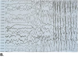
Epilepsy
A disorder characterized by recurrent seizures, diagnosed by EEG.
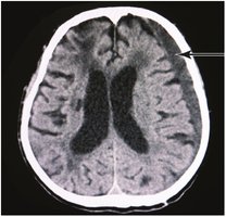
Subdural Hematoma
Bleeding beneath the dura mater, often after trauma.
Migraine
Severe headache often triggered by various factors.

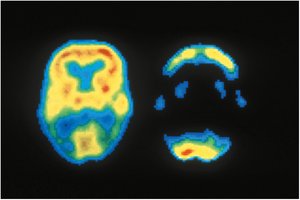
Alzheimer Disease
A progressive neurodegenerative disorder, seen as decreased metabolic activity in PET scans.
Spinal Cord Diseases
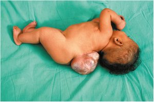
Meningomyelocele
A congenital defect where meninges and spinal cord protrude outside the body.
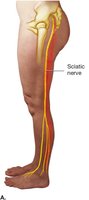
Radiculopathy
Pathology of nerve roots, often due to herniated disk pressing on the sciatic nerve.
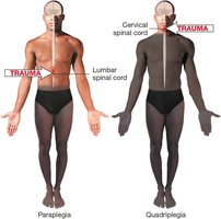
Spinal Cord Injury
Injury level and completeness determine extent of paralysis (paraplegia or quadriplegia).
Nerve Diseases
Bell Palsy
Paralysis of the facial nerve, causing drooping of facial muscles.

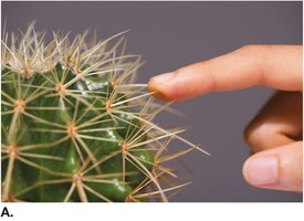
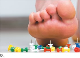
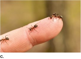
Paresthesias
Abnormal sensations such as burning, tingling, numbness, or crawling feelings.
Laboratory, Diagnostic, and Radiologic Procedures
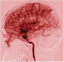
Arteriogram
Imaging of arteries using injected dye to detect blockages or aneurysms.
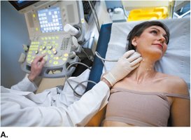
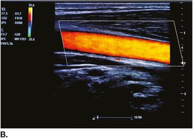
Doppler Carotid Ultrasonography
Ultrasound imaging combined with Doppler technology to assess blood flow in carotid arteries.
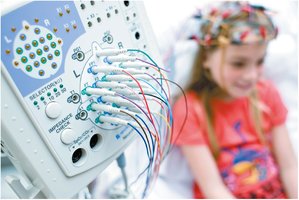
Electroencephalography (EEG)
Recording of electrical impulses of brainwaves to diagnose seizure disorders.
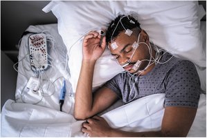
Polysomnography
Sleep study recording EEG, eye movements, muscle activity, heartbeat, and respirations.
Medical Procedures
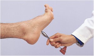
Babinski Sign
A positive Babinski sign indicates abnormal neurological function, seen as extension of the great toe and fanning of other toes.
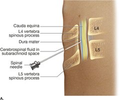
Lumbar Puncture
Procedure to obtain cerebrospinal fluid from the lumbar area for diagnostic purposes.
Summary Table: Divisions of the Nervous System
Division | Main Function | Key Structures |
|---|---|---|
Central Nervous System | Integrates and processes information | Brain, spinal cord |
Peripheral Nervous System | Transmits signals between CNS and body | Cranial and spinal nerves |
Autonomic Nervous System | Controls involuntary functions | Heart, smooth muscle, glands |
Somatic Nervous System | Controls voluntary movements | Skeletal muscles |
Parasympathetic Division | Rest and digest | Acetylcholine |
Sympathetic Division | Fight or flight | Norepinephrine |
Key Equations and Concepts
Neuronal Transmission:
Reflex Arc:
Conclusion
This study guide provides a comprehensive overview of the nervous system, its anatomy, physiology, common diseases, and diagnostic procedures. Understanding these concepts is essential for proficiency in medical terminology and clinical practice related to neurology.
