 Back
BackPeripheral Nervous System, Autonomic Nervous System, and Special Senses: Eye and Ear
Study Guide - Smart Notes
 Tailored notes based on your materials, expanded with key definitions, examples, and context.
Peripheral Nervous System (PNS)
Overview of the PNS
The peripheral nervous system (PNS) consists of all nerves outside the brain and spinal cord. It is responsible for transmitting sensory and motor information between the central nervous system (CNS) and the rest of the body. The PNS is divided into sensory (afferent) and motor (efferent) divisions. The motor division is further subdivided into the somatic (voluntary) and autonomic (involuntary) systems.
Sensory (Afferent): Transmits sensory information from the periphery to the CNS.
Motor (Efferent): Transmits motor commands from the CNS to the periphery.
Somatic: Voluntary control of skeletal muscles.
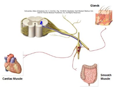
Autonomic: Involuntary control of smooth muscle, cardiac muscle, and glands.
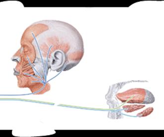
Cranial Nerves
Cranial nerves are twelve pairs of nerves that emerge directly from the brain and brainstem. They are numbered I to XII and may carry sensory, motor, or both types of information. Each nerve has a specific function and area of innervation.
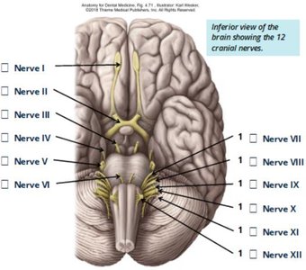
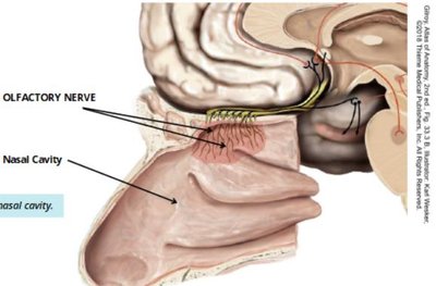
Cranial Nerve I (Olfactory): Sensory only; responsible for the sense of smell.
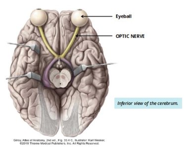
Cranial Nerve II (Optic): Sensory only; responsible for vision.
Cranial Nerve III (Oculomotor): Motor only; controls most of the eye's movements.
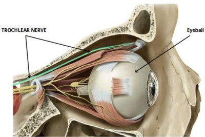
Cranial Nerve IV (Trochlear): Motor only; innervates the superior oblique muscle of the eye.
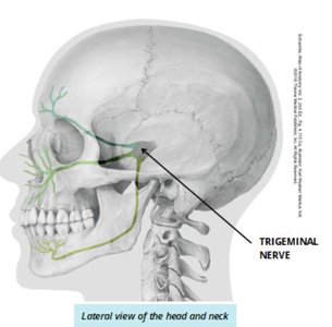
Cranial Nerve V (Trigeminal): Both sensory and motor; provides sensation to the face and motor control to muscles of mastication.
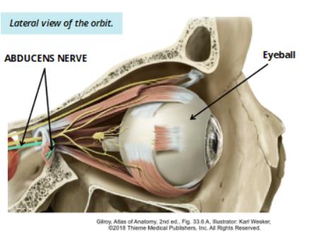
Cranial Nerve VI (Abducens): Motor only; controls the lateral rectus muscle of the eye.
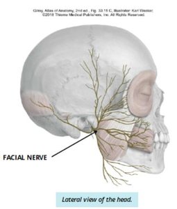
Cranial Nerve VII (Facial): Both sensory and motor; controls muscles of facial expression and taste for the anterior two-thirds of the tongue.
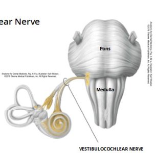
Cranial Nerve VIII (Vestibulocochlear): Sensory only; responsible for hearing and equilibrium.
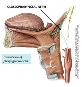
Cranial Nerve IX (Glossopharyngeal): Both sensory and motor; taste from the posterior one-third of the tongue and motor to pharyngeal muscles.
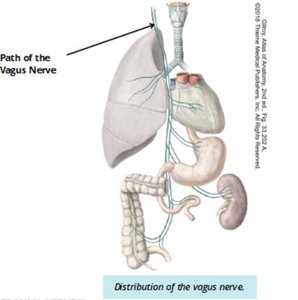
Cranial Nerve X (Vagus): Both sensory and motor; innervates thoracic and abdominal organs, and muscles of the larynx and pharynx.
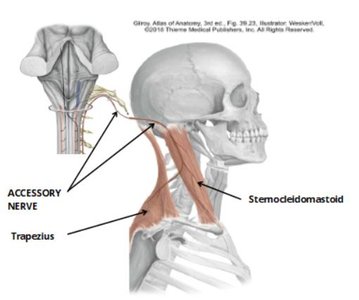
Cranial Nerve XI (Accessory): Motor only; controls the trapezius and sternocleidomastoid muscles.
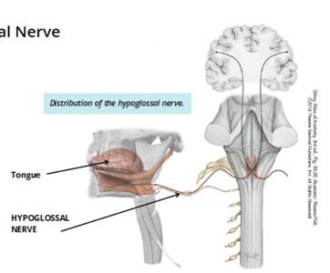
Cranial Nerve XII (Hypoglossal): Motor only; controls muscles of the tongue.
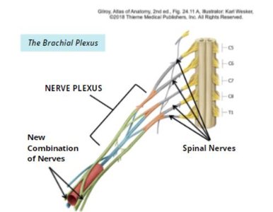
Spinal Nerves and Plexuses
Spinal nerves emerge from the spinal cord and are named according to their region and level. There are 31 pairs of spinal nerves, each formed by the union of sensory (posterior) and motor (anterior) roots. After exiting the vertebral column, anterior root fibers form nerve plexuses, which are networks that redistribute fibers to supply the limbs.
Autonomic Nervous System (ANS)
Overview and Organization
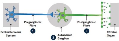
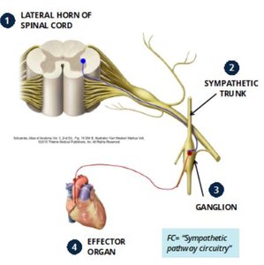
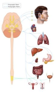
The autonomic nervous system (ANS) is a subdivision of the PNS responsible for involuntary control of smooth muscle, cardiac muscle, and glands. It is organized as a two-neuron pathway: a preganglionic neuron (cell body in CNS) synapses with a postganglionic neuron (cell body in PNS) in an autonomic ganglion.
Divisions of the ANS
Sympathetic Nervous System: Originates from spinal cord segments T1-L2 (thoracolumbar division); responsible for the 'fight or flight' response; has a widespread effect.
Parasympathetic Nervous System: Originates from the brainstem and spinal cord segments S2-S4 (craniosacral division); responsible for 'rest and digest' activities; effects are more localized.
Feature | Sympathetic | Parasympathetic |
|---|---|---|
Origin | T1-L2 (thoracolumbar) | Brainstem, S2-S4 (craniosacral) |
Response | Fight or Flight | Rest and Digest |
Effect | Widespread | Localized |
Special Senses: The Eye
Accessory Structures of the Eye
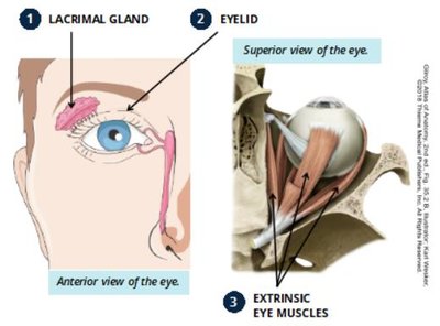
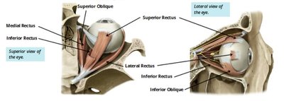
The eye is protected and supported by accessory structures such as the lacrimal gland (produces tears), eyelids (protect and moisten the eye), and extrinsic eye muscles (move the eyeball).
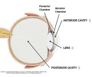
General Features and Cavities of the Eye
The eye is a globe-shaped organ with two main cavities separated by the lens:
Anterior cavity: Contains aqueous humor; divided into anterior and posterior chambers.
Posterior cavity: Contains vitreous humor.
Layers (Tunics) of the Eye
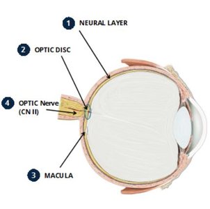
Sensory Tunic (Retina): Innermost layer; contains photoreceptors (rods and cones), optic disc, macula, and optic nerve (CN II).
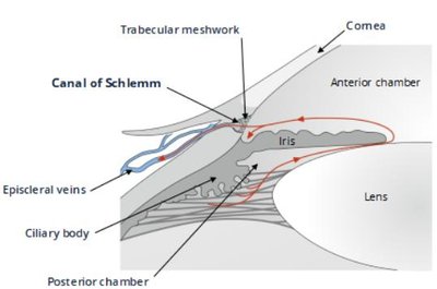
Vascular Tunic (Uveal Tract): Middle layer; includes choroid (blood supply), ciliary body (produces aqueous humor, controls lens shape), and iris (controls pupil size, eye color).
Fibrous Tunic: Outermost layer; consists of the sclera (white of the eye, muscle attachment) and cornea (transparent, allows light entry).
Flow of Aqueous Humor
Aqueous humor is produced by the ciliary body, flows from the posterior chamber through the pupil into the anterior chamber, and drains via the trabecular meshwork and Schlemm's canal into the venous system. Blockage can lead to increased intraocular pressure (glaucoma).
Special Senses: Hearing and Equilibrium
Structure of the Ear
The ear is divided into three main sections:
External Ear: Collects sound waves and directs them to the tympanic membrane (eardrum).
Middle Ear: Contains ossicles (malleus, incus, stapes) that transmit vibrations to the inner ear.
Inner Ear: Contains the cochlea (hearing) and vestibular apparatus (equilibrium).
Inner Ear: Cochlea and Vestibular Apparatus
Cochlea: Spiral-shaped organ containing the organ of Corti, which has hair cells that convert sound vibrations into nerve impulses (transmitted by CN VIII).
Vestibular Apparatus: Includes semicircular canals (detect rotational movement), utricle, and saccule (detect positional movement).
Summary Table: Cranial Nerves and Functions
Nerve | Type | Main Function(s) |
|---|---|---|
Olfactory (I) | Sensory | Smell |
Optic (II) | Sensory | Vision |
Oculomotor (III) | Motor | Eye movement |
Trochlear (IV) | Motor | Eye movement |
Trigeminal (V) | Both | Facial sensation, mastication |
Abducens (VI) | Motor | Eye movement |
Facial (VII) | Both | Facial expression, taste (anterior 2/3 tongue) |
Vestibulocochlear (VIII) | Sensory | Hearing, equilibrium |
Glossopharyngeal (IX) | Both | Taste (posterior 1/3 tongue), pharynx |
Vagus (X) | Both | Thoracic/abdominal organs, larynx/pharynx |
Accessory (XI) | Motor | Sternocleidomastoid, trapezius |
Hypoglossal (XII) | Motor | Tongue muscles |
Additional info: This guide provides foundational knowledge for understanding the structure and function of the peripheral nervous system, autonomic nervous system, and the special senses of vision and hearing, as required for medical terminology and anatomy courses.
