 Back
BackPeripheral Nervous System, Autonomic Nervous System, and Special Senses: Eye and Ear
Study Guide - Smart Notes
 Tailored notes based on your materials, expanded with key definitions, examples, and context.
Peripheral Nervous System (PNS)
Overview of the PNS
The peripheral nervous system (PNS) consists of all nerves outside the brain and spinal cord. It includes both cranial nerves and spinal nerves, which connect the central nervous system (CNS) to limbs and organs.
Sensory (Afferent) Division: Transmits sensory information from the periphery to the CNS.
Motor (Efferent) Division: Transmits motor commands from the CNS to the periphery.
Somatic: Voluntary control of skeletal muscles.
Autonomic: Involuntary control of smooth muscle, cardiac muscle, and glands.
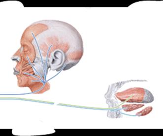
Cranial Nerves
There are 12 pairs of cranial nerves, emerging from the brain and brainstem. They are numbered with Roman numerals I–XII and may carry sensory, motor, or both types of information.
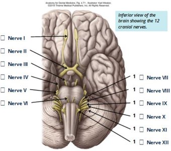
Cranial Nerve | Type | Main Function(s) |
|---|---|---|
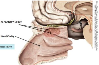
I. Olfactory | Sensory | Smell |
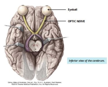
II. Optic | Sensory | Vision |
III. Oculomotor | Motor | Eye movement |
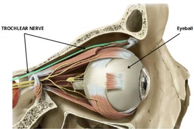
IV. Trochlear | Motor | Eye movement |
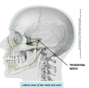
V. Trigeminal | Both | Sensory: Face, Motor: Mastication |
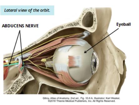
VI. Abducens | Motor | Eye movement |
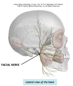
VII. Facial | Both | Sensory: Taste (anterior 2/3 tongue), Motor: Facial expression |
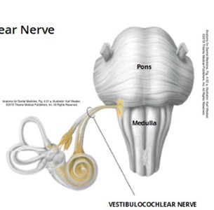
VIII. Vestibulocochlear | Sensory | Hearing, equilibrium |
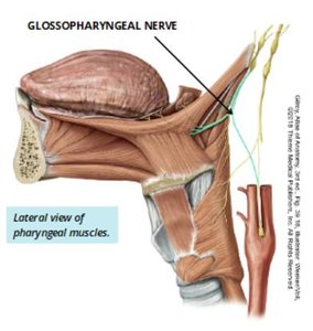
IX. Glossopharyngeal | Both | Sensory: Taste (posterior 1/3 tongue), Motor: Pharynx |
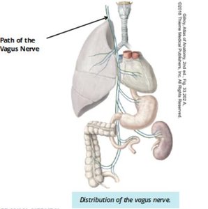
X. Vagus | Both | Sensory/Motor: Thoracic and abdominal organs |
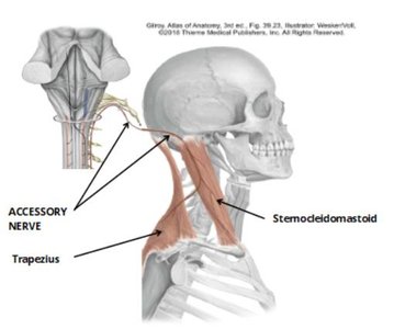
XI. Accessory | Motor | Trapezius, sternocleidomastoid |
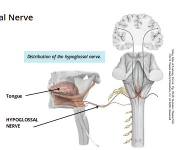
XII. Hypoglossal | Motor | Tongue muscles |
Selected Cranial Nerves and Their Functions
Olfactory Nerve (I): Sensory for smell.
Optic Nerve (II): Sensory for vision.
Oculomotor (III), Trochlear (IV), Abducens (VI): Motor nerves controlling eye movement.
Trigeminal Nerve (V): Sensory for face, motor for mastication.
Facial Nerve (VII): Sensory for taste (anterior 2/3 tongue), motor for facial expression.
Vestibulocochlear Nerve (VIII): Sensory for hearing and equilibrium.
Glossopharyngeal Nerve (IX): Sensory for taste (posterior 1/3 tongue), motor for pharynx.
Vagus Nerve (X): Sensory and motor for thoracic and abdominal organs.
Accessory Nerve (XI): Motor for trapezius and sternocleidomastoid muscles.
Hypoglossal Nerve (XII): Motor for tongue muscles.
Spinal Nerves
There are 31 pairs of spinal nerves, each formed by the union of a posterior (sensory) and anterior (motor) root. They are named according to their region and level of emergence from the spinal cord.
Cervical nerves (C1–C8): Emerge above their corresponding vertebrae, except C8 (between C7 and T1).
Thoracic, lumbar, sacral, and coccygeal nerves: Emerge below their corresponding vertebrae.
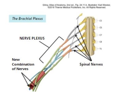
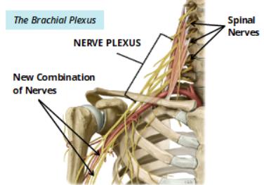
Nerve Plexuses
After exiting the vertebral column, anterior rami of spinal nerves form plexuses (networks) that innervate limbs. Major plexuses include the cervical, brachial, lumbar, and sacral plexuses.
Autonomic Nervous System (ANS)
Overview and Organization
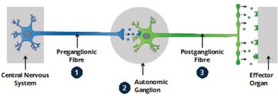
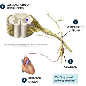
The autonomic nervous system (ANS) is a subdivision of the PNS responsible for involuntary control of smooth muscle, cardiac muscle, and glands. It is organized as a two-neuron pathway:
Preganglionic neuron: Cell body in the CNS, axon projects to an autonomic ganglion.
Autonomic ganglion: Site of synapse between pre- and postganglionic neurons.
Postganglionic neuron: Cell body in the ganglion, axon projects to the effector organ.
Divisions of the ANS
Sympathetic Nervous System: Originates from T1–L2 (thoracolumbar division), responsible for 'fight or flight' responses, with widespread effects.
Parasympathetic Nervous System: Originates from the brainstem and S2–S4 (craniosacral division), responsible for 'rest and digest' responses, with more localized effects.
Feature | Sympathetic | Parasympathetic |
|---|---|---|
Origin | T1–L2 (thoracolumbar) | Brainstem, S2–S4 (craniosacral) |
Response | Fight or Flight | Rest and Digest |
Effect | Widespread | Localized |
Sympathetic Pathways
Preganglionic axons exit the spinal cord via the anterior root, enter the sympathetic trunk, and synapse in ganglia.
Postganglionic axons travel to effector organs at the same or different vertebral levels.
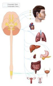
Parasympathetic Pathways
Preganglionic neurons originate in the brainstem or sacral spinal cord, synapse in ganglia near or within target organs.
Postganglionic neurons innervate head (CN III, VII, IX), thorax and upper abdomen (CN X), and pelvic organs (S2–S4).
Special Senses: The Eye
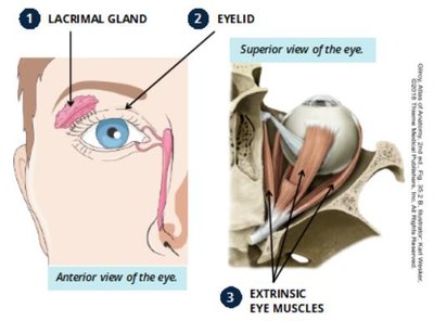
Accessory Structures of the Eye
Accessory structures protect and support the eyeball:
Lacrimal gland: Produces tears to moisten and clean the cornea.
Eyelid: Contains muscles for opening/closing and glands for lubrication.
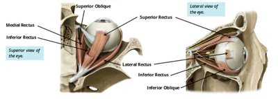
Extrinsic eye muscles: Six muscles control eye movement.
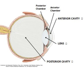
Cavities of the Eye
Anterior cavity: Contains aqueous humor, divided into anterior and posterior chambers.
Lens: Separates anterior and posterior cavities.
Posterior cavity: Contains vitreous humor.
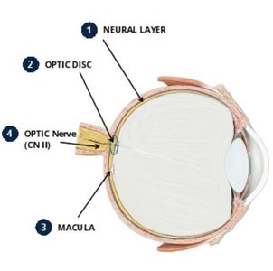
Layers of the Eye (Tunics)
Sensory tunic (Retina): Innermost layer, contains photoreceptors (rods and cones), optic disc, macula, and optic nerve.
Vascular tunic (Uveal tract): Middle layer, includes choroid (blood supply), ciliary body (aqueous humor production, lens shape), and iris (pupil size, eye color).
Fibrous tunic: Outermost layer, includes sclera (white of the eye, muscle attachment) and cornea (transparent, light entry).
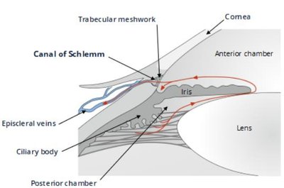
Flow of Aqueous Humor
Aqueous humor is produced by the ciliary body, flows from the posterior chamber through the pupil into the anterior chamber, and drains via the trabecular meshwork and Schlemm’s canal into the venous system.
Special Senses: Hearing and Equilibrium (The Ear)
Structure of the Ear
The ear is divided into three main sections:
External ear: Collects sound waves and directs them to the tympanic membrane (eardrum).
Middle ear: Contains ossicles (malleus, incus, stapes) that transmit vibrations to the inner ear.
Inner ear: Contains receptors for hearing (cochlea) and equilibrium (vestibular apparatus).
Inner Ear: Labyrinths and Receptors
Labyrinths: The bony labyrinth contains the membranous labyrinth. The cochlea (hearing) and vestibular apparatus (equilibrium) are filled with fluids (perilymph and endolymph).
Vestibular apparatus: Semicircular canals detect rotational movement; utricle and saccule detect positional movement.
Cochlea: Contains the organ of Corti, where hair cells transduce sound vibrations into nerve impulses sent via the vestibulocochlear nerve (CN VIII).
Summary Table: Cranial Nerves and Functions
Number | Name | Type | Main Function(s) |
|---|---|---|---|
I | Olfactory | Sensory | Smell |
II | Optic | Sensory | Vision |
III | Oculomotor | Motor | Eye movement |
IV | Trochlear | Motor | Eye movement |
V | Trigeminal | Both | Facial sensation, mastication |
VI | Abducens | Motor | Eye movement |
VII | Facial | Both | Taste, facial expression |
VIII | Vestibulocochlear | Sensory | Hearing, balance |
IX | Glossopharyngeal | Both | Taste, pharynx |
X | Vagus | Both | Viscera, pharynx, larynx |
XI | Accessory | Motor | Neck muscles |
XII | Hypoglossal | Motor | Tongue muscles |
Additional info: This guide provides foundational knowledge for understanding the structure and function of the peripheral nervous system, autonomic nervous system, and the special senses of vision and hearing, as required for medical terminology and anatomy courses.
