 Back
Pulmonology and the Respiratory System: Medical Terminology Study Guide
Study Guide - Smart Notes
 Tailored notes based on your materials, expanded with key definitions, examples, and context.
Pulmonology: Introduction to the Respiratory System
Definition and Scope
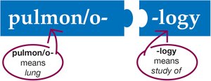
Pulmonology is the medical specialty focused on the anatomy, physiology, and diseases of the respiratory system. Pulmonologists diagnose and treat respiratory disorders using a variety of diagnostic tests, medical and surgical procedures, and pharmacological therapies.
Anatomy of the Respiratory System
Overview of Respiratory Structures
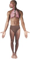
The respiratory system consists of the lungs and a series of connected structures that form the pathway for air to enter and exit the body. It is divided into the upper and lower respiratory tracts.
Upper Respiratory System
Nose and Nasal Cavity
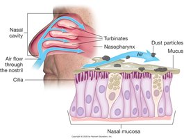
Nose: The entry point for air; contains the nasal cavity, divided by the septum.
Turbinates: Three bony projections that slow airflow, allowing air to be warmed and moistened.
Nasal mucosa: Lines the cavity, humidifies air, and produces mucus to trap particles.
Cilia: Hair-like structures that move mucus and debris out of the nasal passages.
Pharynx
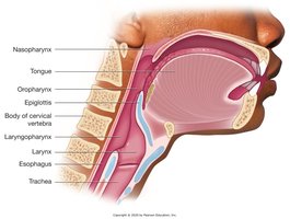
Pharynx: Common passageway for air and food; divided into nasopharynx, oropharynx, and laryngopharynx.
Lower Respiratory System
Larynx
Larynx (voice box): Contains vocal cords; remains open during breathing and closes during swallowing to prevent aspiration.
Trachea and Bronchial Tree
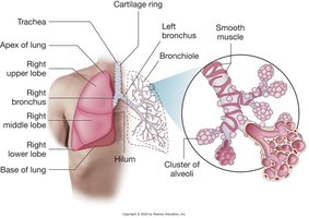
Trachea (windpipe): Supported by C-shaped cartilage rings; divides into right and left primary bronchi.
Bronchi: Enter the lungs and branch into smaller bronchioles; supported by cartilage.
Bronchioles: Smallest airways, with smooth muscle walls and no cartilage; end in alveoli.
Cilia: Line the bronchial tree to move mucus and debris upward.
Lungs and Alveoli
Lungs: Spongy, air-filled organs; right lung has three lobes, left lung has two lobes.
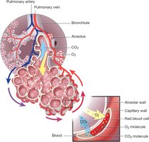
Alveoli: Microscopic air sacs where gas exchange occurs; secrete surfactant to prevent collapse.
Pulmonary parenchyma: Functional tissue of the lungs, mainly alveoli.
Thorax and Pleura
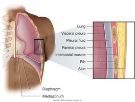
Thorax: Area between the neck and diaphragm; contains the lungs, heart, and other structures.
Pleura: Double-layered serous membrane surrounding each lung; secretes pleural fluid for lubrication.
Mediastinum: Central compartment between the lungs containing the heart, trachea, and esophagus.
Diaphragm: Muscular partition separating thoracic and abdominal cavities; primary muscle of respiration.
Physiology of Respiration
Breathing Mechanism
Inhalation (Inspiration): Diaphragm contracts and moves downward, intercostal muscles lift ribs, thoracic cavity enlarges, and air flows in due to negative pressure.
Exhalation (Expiration): Diaphragm relaxes and moves upward, intercostal muscles relax, thoracic cavity shrinks, and air is expelled due to positive pressure.
Control: Respiratory centers in the brainstem regulate rate and depth via the phrenic nerve.

Processes of Respiration
Ventilation: Movement of air in and out of the lungs.
External respiration: Gas exchange between alveoli and blood.
Gas transport: Movement of oxygen and carbon dioxide in the blood.
Internal respiration: Exchange of gases between blood and body cells.
Cellular respiration: Cells use oxygen for metabolism, producing carbon dioxide as waste.
Common Respiratory Diseases
Diseases of the Nose and Pharynx
Upper Respiratory Infection (URI): Caused by bacteria or viruses; spreads via droplets or contact.

Diseases of the Lungs
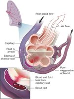
Acute Respiratory Distress Syndrome (ARDS): Alveoli fill with fluid, leading to poor oxygenation and blood clots.
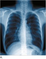
Atelectasis: Collapse of part or all of a lung, seen as a hazy area on chest x-ray.
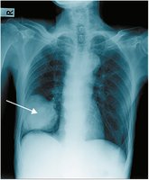
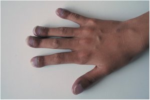
Cystic Fibrosis: Genetic disorder causing thick mucus, cyanosis, and clubbing of fingers due to chronic hypoxia.
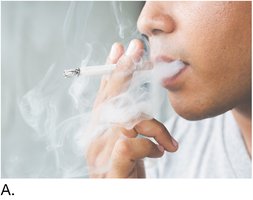
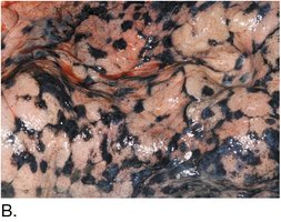
Effects of Smoking: Inhalation of tar leads to black deposits in lung tissue, increasing risk of respiratory diseases.
Pneumonia: Infection causing consolidation (dense area) in the lung, visible on x-ray.
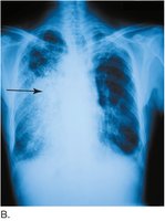
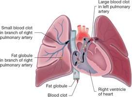
Pulmonary Embolus: Blood clot or fat globule blocks a pulmonary artery, preventing blood from reaching alveoli and causing collapse in that lung area.
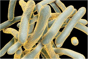
Tuberculosis: Caused by Mycobacterium tuberculosis, a bacterium with a waxy coating that resists treatment.
Diseases of Respiration
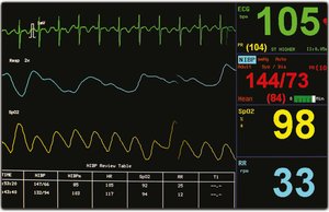
Tachypnea: Abnormally rapid breathing; normal adult rate is 12–20 breaths per minute.
Laboratory Tests and Diagnostic Procedures
Pulse Oximetry
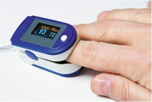
Pulse Oximeter: Measures oxygen saturation of hemoglobin and pulse rate; quick, noninvasive assessment.
Pulmonary Function Tests (PFTs)
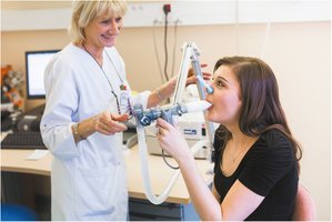
Pulmonary Function Test: Measures lung volumes and capacities; used to monitor diseases like cystic fibrosis.
Sleep Studies
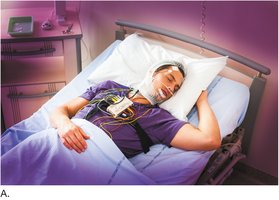
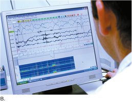
Polysomnography (Sleep Study): Monitors breathing and other parameters during sleep to diagnose disorders like sleep apnea.
Radiologic Procedures
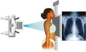
Chest X-Ray (PA View): Standard imaging to assess lung fields, heart size, and detect abnormalities.
Medical Procedures
Auscultation and Percussion
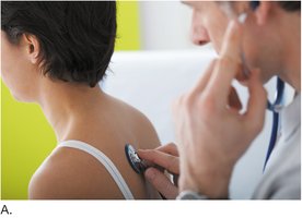
Auscultation: Listening to breath sounds with a stethoscope to detect abnormalities.
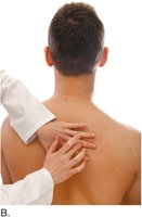
Percussion: Tapping on the chest to identify areas of dullness or resonance.
Chest Percussion Therapy
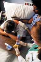
Chest Percussion Therapy: Used to loosen thick mucus in conditions like cystic fibrosis, facilitating expectoration.
Positive Airway Pressure Devices
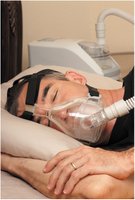
CPAP Machine: Provides continuous positive airway pressure to keep airways open during sleep, commonly used for sleep apnea.
Endotracheal Intubation
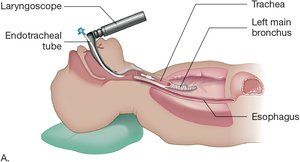
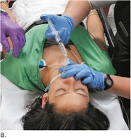
Endotracheal Intubation: Placement of a tube into the trachea to maintain an open airway or deliver drugs/ventilation.
Incentive Spirometry
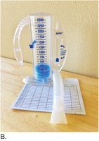
Incentive Spirometer: Device encouraging deep breathing to prevent atelectasis postoperatively; measures inspired air volume.

Oxygen Therapy
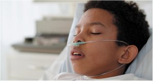
Nasal Cannula: Delivers supplemental oxygen via prongs in the nostrils; up to 40% oxygen concentration.
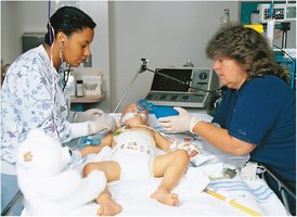
Oxygen Therapy with Respirator: Used in critical care to ensure adequate oxygenation, especially in infants or severe respiratory distress.
Drugs and Surgical Procedures
Inhalers and Smoking Cessation
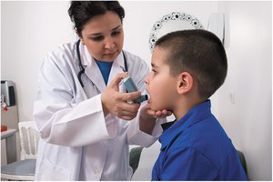
Metered-Dose Inhaler: Delivers a measured dose of bronchodilator or corticosteroid directly to the lungs.
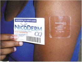
Smoking Cessation Drug: Nicotine patch provides a controlled dose of nicotine to help patients quit smoking.
Surgical Procedures
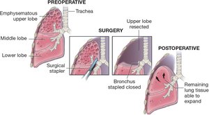
Lobectomy: Surgical removal of a lung lobe, often for emphysema or cancer; remaining lung tissue expands to fill the space.
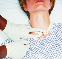
Tracheostomy: Surgical creation of an opening in the trachea to facilitate breathing; tube is secured and area is kept clean to prevent infection.
Additional info: This guide covers the essential terminology, anatomy, physiology, diseases, diagnostic procedures, and treatments relevant to the respiratory system, as outlined in a typical Medical Terminology college course.
