Back
BackStructural Organization of the Human Body: Medical Terminology Study Guide
Study Guide - Smart Notes
 Tailored notes based on your materials, expanded with key definitions, examples, and context.
Whole Body Terms and Structural Organization
Levels of Structural Organization
The human body is organized into hierarchical levels, each with increasing complexity. Understanding these levels is fundamental to medical terminology and anatomy.
Cells: The smallest and most numerous structural units of the body. Cells carry out essential life functions.
Tissues: Groups of similar cells working together to perform specialized functions. Four main types: connective, epithelial, muscle, and nervous.
Organs: Structures composed of two or more types of tissues, arranged to perform specific functions.
Body Systems: Groups of organs working together to perform complex functions necessary for life. Examples include the integumentary, skeletal, muscular, nervous, cardiovascular, respiratory, digestive, urinary, special senses, endocrine, blood/lymphatic, and reproductive systems.
Example: The digestive system includes organs such as the stomach and intestines, which are made up of all four tissue types.
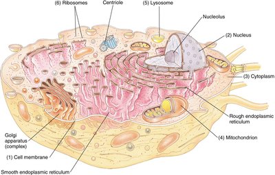
Component Parts of a Cell
Cells contain specialized structures called organelles, each with distinct functions vital for cellular activity.
Cell membrane: The outer boundary of the cell, controlling entry and exit of substances.
Nucleus: Contains genetic material (DNA) and controls cellular activities.
Cytoplasm: Gel-like substance where organelles are suspended.
Mitochondrion: The powerhouse of the cell, responsible for energy production.
Rough and Smooth Endoplasmic Reticulum: Involved in protein and lipid synthesis.
Golgi apparatus: Processes and packages proteins and lipids.
Lysosome: Contains digestive enzymes for breaking down waste.
Ribosomes: Sites of protein synthesis.
Terms Relating to Cells
Medical terminology often uses prefixes and suffixes to describe cellular conditions:
Anaplasia: Lack of differentiation or formation.
Aplasia: Absence or failure of development.
Dysplasia: Abnormal growth or development.
Hyperplasia: Excessive cell proliferation.
Hypoplasia: Underdevelopment or below normal growth.
Neoplasia: New, uncontrolled growth (often refers to tumors).
Types of Tissues
The body is composed of four primary tissue types, each with unique functions:
Connective tissue: Supports and binds other tissues (e.g., bone, blood).
Epithelial tissue: Covers internal and external surfaces of organs.
Muscle tissue: Responsible for movement; includes skeletal (attached to bone), smooth (walls of internal organs), and cardiac (heart wall).
Nervous tissue: Transmits electrical impulses for communication.
Example: All four tissue types are present in every body system, such as the digestive system.
Body Planes
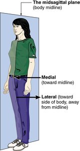
Midsagittal Plane
The midsagittal plane is a vertical line dividing the body into equal right and left halves. It is essential for describing locations and directions in anatomy.
Medial: Toward the midline of the body.
Lateral: Away from the midline, toward the sides.
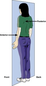
Frontal (Coronal) Plane
The frontal or coronal plane is a vertical plane dividing the body into anterior (front) and posterior (back) portions.
Anterior: Toward the front of the body.
Posterior: Toward the back of the body.
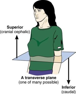
Transverse Plane
The transverse plane is a horizontal line dividing the body into superior (upper) and inferior (lower) portions.
Superior (cranial, cephalic): Above or toward the head.
Inferior (caudal): Below or toward the feet.
Body Regions and Abdominal Quadrants
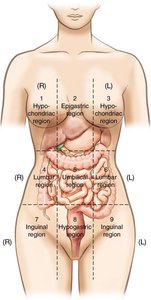
Nine Abdominal Regions
The abdomen is divided into nine regions for precise anatomical and clinical reference. These regions are identified from left to right, top to bottom.
Right and left hypochondriac regions
Epigastric region
Right and left lumbar regions
Umbilical region
Right and left inguinal regions
Hypogastric region
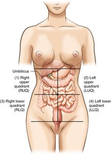
Abdominal Quadrants
The abdomen is also divided into four quadrants, using the umbilicus (navel) as the landmark:
Right Upper Quadrant (RUQ)
Left Upper Quadrant (LUQ)
Right Lower Quadrant (RLQ)
Left Lower Quadrant (LLQ)
McBurney’s point: A clinical landmark in the RLQ, often used to diagnose appendicitis.
Munro’s point: Another clinical landmark in the LLQ.
Body Cavities
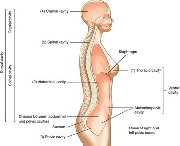
Major Body Cavities
The body contains two main cavities, each housing specific organs:
Ventral cavity: Includes the thoracic, abdominal, and pelvic cavities.
Dorsal cavity: Includes the cranial and spinal cavities.
The diaphragm separates the thoracic and abdominal cavities, playing a crucial role in respiration.
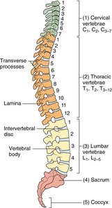
Divisions of the Back
Vertebral Column Regions
The vertebral column is divided into five regions, each with a specific number of vertebrae:
Cervical vertebrae (C1–C7): Neck region
Thoracic vertebrae (T1–T12): Upper and mid-back
Lumbar vertebrae (L1–L5): Lower back
Sacrum: Five fused vertebrae
Coccyx: Four fused vertebrae (tailbone)
Directional Terms
Common Directional Terms
Directional terms are used to describe the location of structures relative to other parts of the body.
Superficial: Near the surface
Deep: Away from the surface
Anterior (ventral): Front
Posterior (dorsal): Back
Superior (cranial): Above
Inferior (caudal): Below
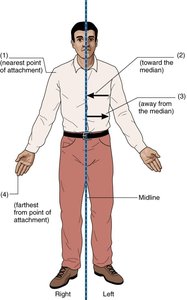
Additional Directional Terms
Further terms help specify positions and movements:
Medial: Toward the midline
Lateral: Away from the midline
Distal: Farther from the point of attachment
Proximal: Nearer to the point of attachment
Supine: Lying face up
Prone: Lying face down
Supination: Turning the palm upward
Pronation: Turning the palm downward
Plantar: Sole of the foot
Dorsal: Back of the body or top of the foot