 Back
BackStudy Guide: Medical Terminology – Nervous System
Study Guide - Smart Notes
 Tailored notes based on your materials, expanded with key definitions, examples, and context.
Nervous System Overview
Divisions of the Nervous System
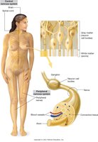
The nervous system is a complex network responsible for coordinating bodily functions and responding to internal and external stimuli. It is divided into two main parts: the Central Nervous System (CNS) and the Peripheral Nervous System (PNS).
CNS: Includes the brain and spinal cord, protected by the skull and vertebral column.
PNS: Comprises nerves branching from the CNS, including 12 pairs of cranial nerves and 31 pairs of spinal nerves.
Key Functions
Receives and processes sensory information.
Coordinates motor responses.
Maintains homeostasis through involuntary and voluntary actions.
Tissues of the Nervous System
Principal Tissue Types
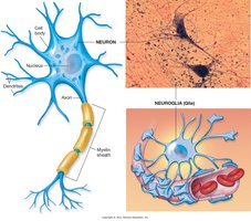
The nervous system is composed of two main tissue types: neurons and neuroglia.
Neurons: Structural and functional units that conduct impulses.
Neuroglia: Supporting cells that provide structural and metabolic support.
Types of Neurons
Motor Neurons: Transmit impulses from CNS to muscles and glands (efferent).
Sensory Neurons: Carry impulses from sensory receptors to CNS (afferent).
Interneurons: Mediate impulses between sensory and motor neurons, located entirely within CNS.
Neuron Structure
Cell Body: Contains nucleus and metabolic machinery.
Axon: Long process transmitting impulses away from cell body; often myelinated for faster conduction.
Dendrites: Short, branched processes transmitting impulses toward cell body.
Nerve Fibers, Nerves, and Tracts
Nerve Fibers
Myelinated fibers: Have a myelin sheath and neurilemma (Schwann cells) for rapid impulse transmission.
Unmyelinated fibers: Lack myelin, slower transmission.
Nerves and Tracts
Nerves: Bundles of nerve fibers outside CNS; classified as afferent (sensory) or efferent (motor).
Tracts: Bundles of nerve fibers within CNS, grouped by origin, function, and termination.
Transmission of Nerve Impulses
All-or-None Principle
No transmission occurs until stimulus reaches threshold.
Once threshold is reached, maximum impulse is produced.
Synapse and Neurotransmitters
Impulse transmitted across synaptic cleft via neurotransmitters.
Synaptic cleft separates axon terminals from dendrites or muscle end plates.
Central Nervous System
Brain and Spinal Cord
Gray Matter: Unsheathed cell bodies and dendrites.
White Matter: Myelinated nerve fibers.
Embryonic Development
Neural tube forms CNS at 3-4 weeks.
Brain waves measurable at 6 weeks.
Regulatory functions begin at 28 weeks; rapid growth until age 4.
Brain Structure and Function
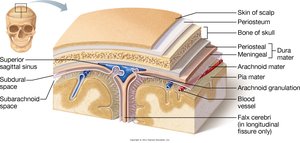
Meninges
The brain is protected by three membranes called meninges:
Dura mater: Outermost, tough layer.
Arachnoid: Middle, web-like layer.
Pia mater: Innermost, delicate layer.
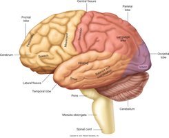
Major Brain Structures
Cerebrum: Largest part, controls sensory and motor activity, memory, emotions, consciousness.
Cerebellum: Coordinates movement and maintains posture.
Diencephalon: Includes thalamus, hypothalamus, epithalamus; regulates sensory relay, emotions, autonomic functions.
Brainstem: Includes midbrain, pons, medulla oblongata; controls vital functions and cranial nerve innervation.
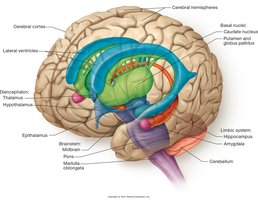
Cerebrum and Lobes
Frontal lobe: Motor area, personality, speech.
Parietal lobe: Sensory input, language interpretation.
Temporal lobe: Hearing, smell, language input.
Occipital lobe: Vision processing.
Hippocampus
Part of limbic system, involved in emotions and memory.
Atrophy linked to Alzheimer disease, depression, schizophrenia.
Cerebellum
Second largest brain part, coordinates voluntary/involuntary movements.
Maintains muscle tone and posture.
Diencephalon
Epithalamus: Maintains circadian rhythms, connects limbic system, stimulates melatonin secretion.
Thalamus: Relay center for sensory/motor impulses.
Hypothalamus: Regulates autonomic activity, water balance, metabolism, temperature.
Pituitary gland: Attached to hypothalamus, controls endocrine functions.
Brainstem
Midbrain: Visual and auditory reflexes.
Pons: Links cerebellum and medulla to higher brain areas; motor control.
Medulla oblongata: Controls breathing, heart rate, blood pressure, swallowing, vomiting.
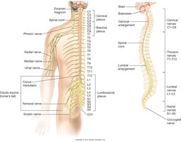
Spinal Cord
Structure and Function
H-shaped gray matter surrounded by white matter.
Conducts sensory input to brain and motor output from brain.
Serves as reflex center.
Key Regions
Conus medullaris: Tapered end between T12 and L1.
Filum terminale: Fibrous thread extending to sacral vertebra.
Cauda equina: Bundle of lumbar, sacral, coccygeal nerves.
Cerebrospinal Fluid (CSF)
Production and Function
Produced by choroid plexuses in brain ventricles.
Cushions brain and spinal cord, supports brain, carries neurotransmitters.
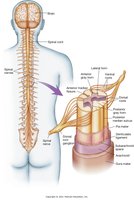
Peripheral Nervous System
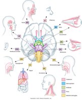
Cranial Nerves
12 pairs, provide sensory and motor functions.
Named for area/function served.
Spinal Nerves
31 pairs, named for vertebral region.
Each nerve has dorsal (sensory) and ventral (motor) roots.
Dorsal rami: Serve muscles/skin of back.
Ventral rami: Serve larger areas including limbs and organs.
Autonomic Nervous System
Overview
The autonomic nervous system (ANS) is part of the PNS and controls involuntary functions such as glandular secretion, blood pressure, and heart rate. 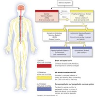
Divisions
Sympathetic: Prepares body for fight-or-flight; increases alertness, heart rate, blood pressure, and metabolic rate.
Parasympathetic: Conserves energy; stimulates digestion, slows heart rate, reduces blood pressure.
Sympathetic Trunk
Chain of ganglia running alongside vertebral column.
Produces widespread innervation.
Parasympathetic Division
Long fibers from cranial and sacral nerves.
Ganglia located near target organs.
Summary Table: Nervous System Divisions
Division | Main Structures | Function |
|---|---|---|
Central Nervous System (CNS) | Brain, Spinal Cord | Integrates and processes information |
Peripheral Nervous System (PNS) | Cranial and Spinal Nerves | Transmits signals between CNS and body |
Autonomic Nervous System (ANS) | Sympathetic & Parasympathetic branches | Controls involuntary functions |
Key Medical Terminology
Afferent: Conducts impulses toward CNS (sensory).
Efferent: Conducts impulses away from CNS (motor).
Ganglia: Clusters of nerve cell bodies outside CNS.
Corpus callosum: Largest brain tract connecting hemispheres.
Myelin sheath: Fatty covering for axons, increases impulse speed.
Additional info:
Embryonic development milestones and clinical relevance of hippocampal atrophy were inferred for completeness.
