 Back
BackStudy Guide: The Endocrine System (ANAT 100 Module 07)
Study Guide - Smart Notes
 Tailored notes based on your materials, expanded with key definitions, examples, and context.
Overview of the Endocrine System
Introduction to Endocrine Glands
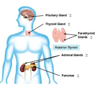
The endocrine system is a network of glands that secrete hormones directly into the bloodstream to regulate various bodily functions. These glands play a crucial role in maintaining homeostasis and influencing other organ systems.
Endocrine glands release hormones into the blood; examples include the pituitary, thyroid, parathyroid, adrenal glands, and pancreas.
Exocrine glands secrete products into ducts (e.g., sweat, salivary glands).
Hormones are chemical messengers that act on target organs to elicit specific responses.
The endocrine system affects the cardiovascular, reproductive, and digestive systems, among others.
Pituitary Gland (Hypophysis)
Anatomical Location and Structure
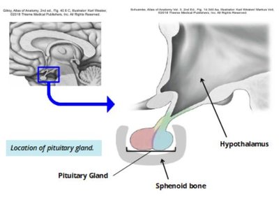
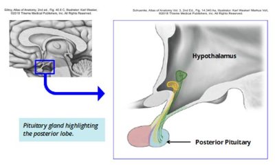
The pituitary gland is a small, pea-sized gland located inferior to the hypothalamus, sitting in the sphenoid bone's concavity. It is often called the 'master gland' because it regulates other endocrine glands.
Physical contact with the hypothalamus allows for nervous and endocrine system communication.
Divided into two lobes: anterior lobe (adenohypophysis) and posterior lobe (neurohypophysis).
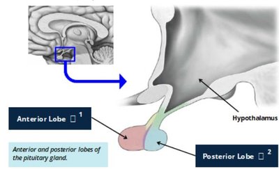
Anterior Lobe (Adenohypophysis)
The anterior lobe is composed of glandular secretory cells and produces seven stimulating hormones, each with specific target organs and functions.
Thyroid Stimulating Hormone (TSH): Stimulates thyroid hormone release.
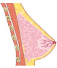
Prolactin (PRL): Induces milk production in mammary glands.
Adrenocorticotropic Hormone (ACTH): Stimulates adrenal gland hormone release.
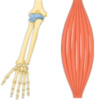
Growth Hormone (GH): Promotes growth in all cells.
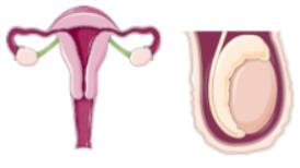
Follicle Stimulating Hormone (FSH) & Luteinizing Hormone (LH): Regulate gonadal function (estrogen, progesterone, testosterone, gamete production).
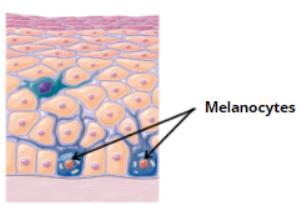
Melanocyte Stimulating Hormone (MSH): Stimulates skin melanocytes to produce pigment.
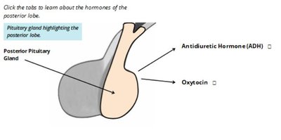
Posterior Lobe (Neurohypophysis)
The posterior lobe is composed of nervous tissue and releases two hormones produced in the hypothalamus.
Antidiuretic Hormone (ADH): Promotes water reabsorption in the kidneys.
Oxytocin: Promotes uterine contractions during childbirth and plays a role in breastfeeding.
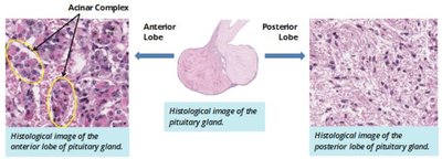
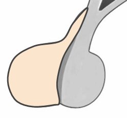
Pituitary Gland Histology
The anterior and posterior lobes differ microscopically. The anterior lobe is made of glandular acini and stains darker, while the posterior lobe is nervous tissue and stains lighter.
Anterior lobe: Glandular acini, darker staining.
Posterior lobe: Nervous and connective tissue, lighter staining.
Thyroid Gland
Anatomical Features
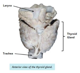
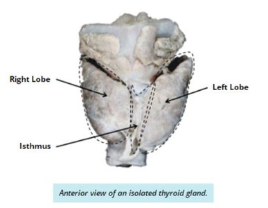
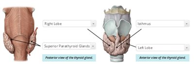
The thyroid gland is located in the neck, anterior to the trachea and inferior to the larynx. It is divided into right and left lobes connected by the isthmus.
Maintains metabolic homeostasis.
Shape: Butterfly-like, with two lobes and a central isthmus.
Histology and Cell Types
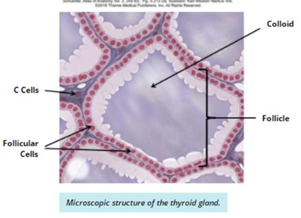
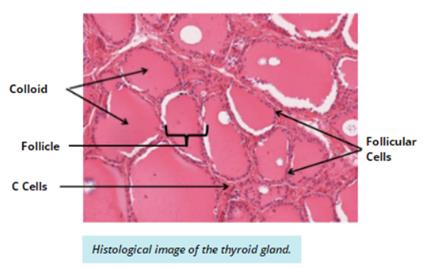
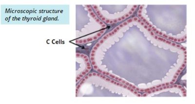
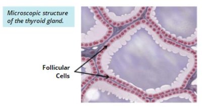
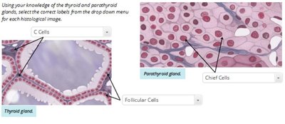
The thyroid gland is composed of follicles lined by simple cuboidal follicular cells, surrounding a central lumen filled with colloid. Parafollicular (C) cells are dispersed between follicles.
Follicular cells: Produce T3 and T4 hormones, increasing metabolism and oxygen consumption.
C cells: Produce calcitonin, lowering blood calcium concentration.
Colloid: Protein-rich fluid secreted by follicular cells.
Case Study: Goitre
A goitre is an enlargement of the thyroid gland, often caused by an imbalance in T3 and T4 hormone levels. Excess TSH from the pituitary can lead to increased T3 and T4, causing goitre.
TSH: Pituitary hormone responsible for stimulating thyroid hormone production.
Parathyroid Glands
Anatomical and Histological Features
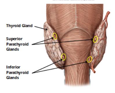
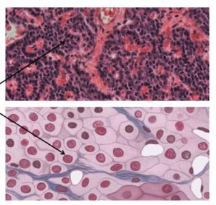
The parathyroid glands are four small oval glands located on the posterior side of the thyroid. They are paired (two superior, two inferior) and contribute to calcium homeostasis.
Principal (chief) cells: Produce parathyroid hormone (PTH), which increases blood calcium levels.
Adrenal Glands
Anatomical Location and Structure
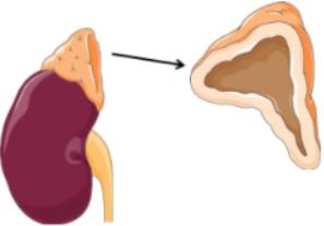
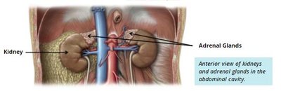
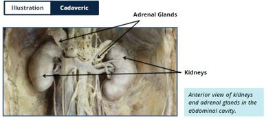
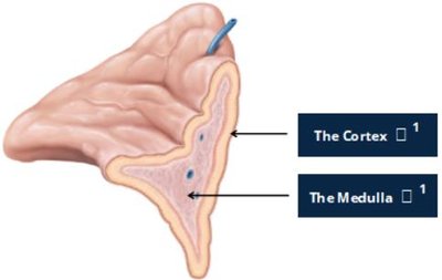
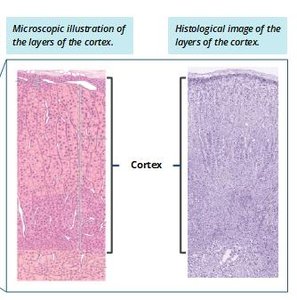
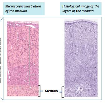
The adrenal glands are pyramid-shaped structures sitting atop each kidney. Each gland is divided into the cortex and medulla, which produce different hormones.
Adrenal cortex: Three layers producing corticosteroids (mineralocorticoids, glucocorticoids, androgens).
Adrenal medulla: Produces epinephrine and norepinephrine for the fight-or-flight response.
Pancreas
Anatomical Location and Dual Function
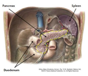
The pancreas is a long, lobular organ located posterior to the stomach, extending from the duodenum to the spleen. It has both endocrine and exocrine functions.
Endocrine function: Regulates blood sugar via hormone secretion.
Exocrine function: Produces digestive enzymes.
Endocrine Cells of the Pancreas
The endocrine cells are organized into pancreatic islets (islets of Langerhans), which contain two main cell types:
Alpha cells: Produce glucagon, increasing blood glucose levels.
Beta cells: Produce insulin, lowering blood glucose levels.
Clinical Application: Type I Diabetes
Type I diabetes is caused by destruction of beta cells, resulting in loss of insulin production. Without insulin, glucose cannot be absorbed, leading to hyperglycemia and associated symptoms.
Treatment: Injection of exogenous insulin.
Summary Table: Major Endocrine Glands and Their Hormones
Gland | Main Hormones | Main Functions |
|---|---|---|
Pituitary (Anterior) | TSH, PRL, ACTH, GH, FSH, LH, MSH | Regulates other glands, growth, reproduction, pigmentation |
Pituitary (Posterior) | ADH, Oxytocin | Water balance, uterine contraction, lactation |
Thyroid | T3, T4, Calcitonin | Metabolism, calcium homeostasis |
Parathyroid | PTH | Increases blood calcium |
Adrenal Cortex | Mineralocorticoids, Glucocorticoids, Androgens | Electrolyte balance, metabolism, sex characteristics |
Adrenal Medulla | Epinephrine, Norepinephrine | Fight-or-flight response |
Pancreas | Insulin, Glucagon | Blood glucose regulation |
Conclusion
The endocrine system is essential for regulating body functions through hormone secretion. Understanding the anatomy, histology, and function of each gland is crucial for medical terminology and clinical practice.
