 Back
BackStudy Guide: The Nervous System (Central Nervous System Focus)
Study Guide - Smart Notes
 Tailored notes based on your materials, expanded with key definitions, examples, and context.
Introduction to the Nervous System
Overview
The nervous system is the body's primary control and communication network, responsible for both voluntary and involuntary actions. It consists of the brain, spinal cord, sensory organs, and all nerves throughout the body. This module focuses on the central nervous system (CNS), its structure, function, and cellular composition.
Organization of the Nervous System
Functions of the Nervous System
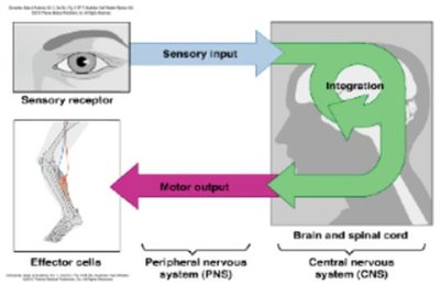
The nervous system operates through three main steps: sensory input, integration, and motor output.
Sensory Input: Gathering information from internal and external environments via sensory receptors.
Integration: Processing and interpreting sensory input to form a complete picture, primarily in the brain and spinal cord.
Motor Output: Sending signals to effector cells (muscles or glands) to produce a response.
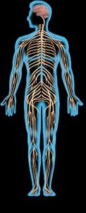
Divisions of the Nervous System
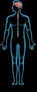
Central Nervous System (CNS): Composed of the brain and spinal cord; acts as the control center.
Peripheral Nervous System (PNS): Includes all nerves outside the CNS, such as cranial and spinal nerves, autonomic nervous system, and special sense organs.
Afferent vs. Efferent Neurons
Afferent (Sensory) Neurons: Carry signals to the CNS from sensory receptors.
Efferent (Motor) Neurons: Carry signals away from the CNS to effector cells.
Similarity: Both are part of the PNS and transmit electrical signals.
Difference: Direction of signal transmission (to vs. from CNS).
Histology of Neural Tissue
Principal Cell Types
Neurons: Excitable cells that generate and transmit electrical signals.
Supporting Cells (Neuroglia/Glia): Non-excitable cells that protect, support, and enhance communication between neurons.
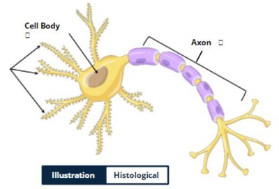
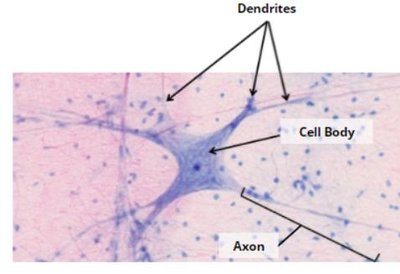
Neuron Structure
Neurons have three main components:
Cell Body (Soma): Contains the nucleus and organelles; transfers signals from dendrites to axon.
Dendrites: Receive signals from other neurons and transmit them to the cell body.
Axon: Single, long process that carries signals from the cell body to axon terminals.
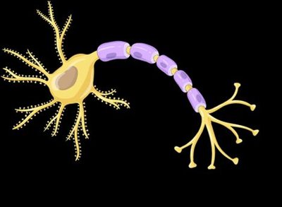
Direction of Nerve Impulse
Signals travel unidirectionally: dendrites → cell body → axon → axon terminals.
Signals are not transmitted in the reverse direction.
Classification of Neurons
Pseudounipolar (Unipolar): One process splits into dendrite and axon; found in general sensory nerves.
Bipolar: One dendrite and one axon; found in special senses (sight, hearing).
Multipolar: Multiple dendrites and one axon; most common, includes motor neurons.
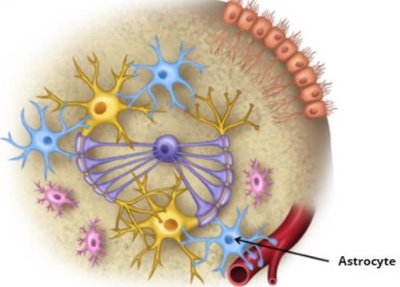
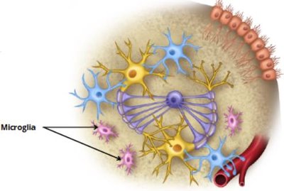
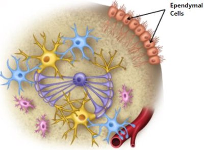
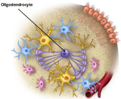
Supporting Cells of the CNS
Astrocytes: Star-shaped, maintain neural tissue integrity, most abundant glial cell.
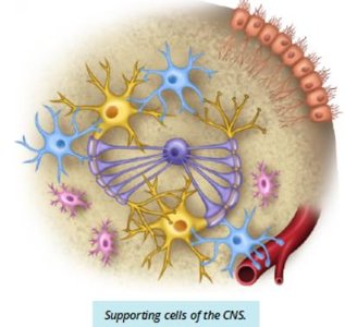
Microglia: Small, phagocytose pathogens and waste, least common.
Ependymal Cells: Cuboidal, line brain and spinal cord cavities, produce and circulate cerebrospinal fluid (CSF).
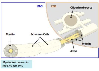
Oligodendrocytes: Myelinate axons in CNS, one cell can myelinate up to 60 axons.
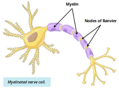
Myelination
Myelin: Fatty substance that insulates axons, increases speed of electrical signal transmission.
Nodes of Ranvier: Unmyelinated gaps between myelinated regions, facilitate rapid signal conduction.
Demyelination: Diseases like multiple sclerosis involve loss of myelin, leading to neurological symptoms.
Supporting Cells of the PNS
Schwann Cells: Myelinate axons in the PNS; each cell myelinates only one segment of one axon.
Comparison: Oligodendrocytes (CNS) vs. Schwann cells (PNS) in myelination.
Central Nervous System – The Brain
Major Regions of the Brain
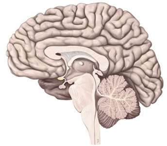
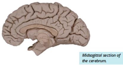
Cerebrum: Includes telencephalon and diencephalon; responsible for sensory interpretation, motor output, and decision making.
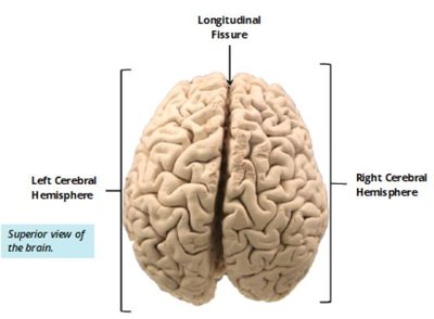
Cerebral Hemispheres: Right and left hemispheres separated by the longitudinal fissure.
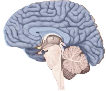
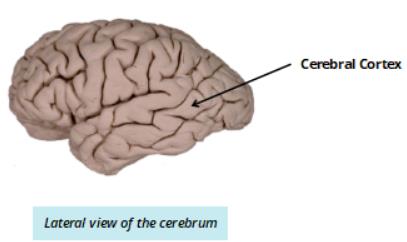
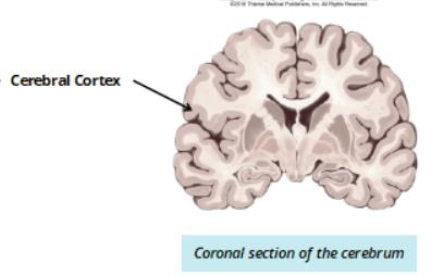
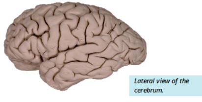
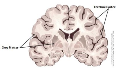
Cerebral Cortex: Outer layer, dark grey in coronal section, site of higher brain functions.
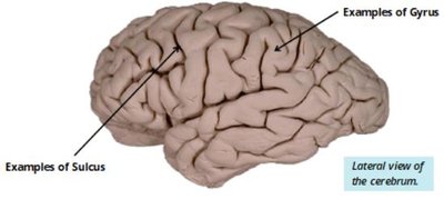
Sulci and Gyri
Sulci: Grooves/fissures that increase surface area and divide brain regions.
Gyri: Raised areas between sulci, associated with specific functions.
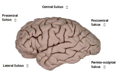
Major Sulci of the Cerebrum
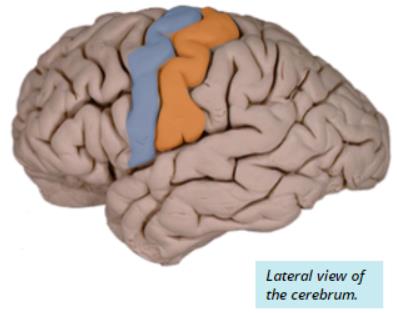
Precentral Sulcus: Anterior border of motor cortex.
Central Sulcus: Divides frontal and parietal lobes; separates motor and sensory cortices.
Postcentral Sulcus: Posterior border of sensory cortex.
Parieto-occipital Sulcus: Divides parietal and occipital lobes.
Lateral Sulcus: Divides temporal lobe from frontal and parietal lobes.
Major Gyri of the Cerebrum
Precentral Gyrus: Motor cortex, sends motor output signals (anterior to central sulcus).
Postcentral Gyrus: Somatosensory cortex, receives sensory input (posterior to central sulcus).
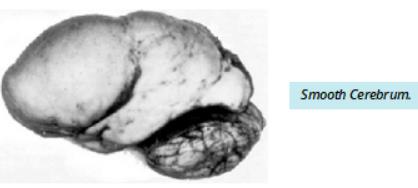
Clinical Application: Lissencephaly (Smooth Brain Syndrome)
Definition: Lack of development of sulci and gyri.
Functional Impact: Reduced surface area, limited mental capacity, severe intellectual disability, slowed physical development, often incompatible with life beyond 10 years.
Lobes of the Cerebrum
Frontal Lobe: Anterior to central sulcus; involved in motor function, problem solving, and speech.
Parietal Lobe: Between central and parieto-occipital sulcus; processes sensory information.
Occipital Lobe: Posterior to parieto-occipital sulcus; responsible for visual processing.
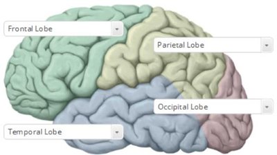
Temporal Lobe: Inferior to lateral sulcus; involved in auditory processing and memory.
Grey and White Matter of the Brain
Grey Matter: Contains neuron cell bodies, interneurons, and glial cells; found in cerebral cortex and inner brain areas.
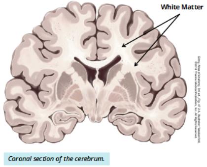
White Matter: Bundles of nerve fibers and axons; found in inner regions of cerebrum. The corpus callosum connects the two hemispheres.
White Matter Tracts
Association Fibres: Communication within one hemisphere.
Commissural Fibres: Communication between hemispheres (e.g., corpus callosum).
Projection Fibres: Communication between different CNS levels (brain and spinal cord).
Central Nervous System – Spinal Cord
Spinal Cord Structure and Function
Located in the vertebral canal, supported by the vertebral column.
Facilitates communication between CNS and PNS via spinal nerves.
Spinal nerves supply sensory and motor innervation to regions near their exit point.
External Topography
Begins at foramen magnum, extends to L2 (conus medullaris).
Cauda equina: Axons emerging below L2, resembling a horse's tail.
Filum terminale: Strand of pia mater anchoring spinal cord to sacrum and coccyx.
Regions of the Spinal Cord
Cervical: Continuous with brainstem, housed in neck vertebrae.
Thoracic: Located in upper back vertebrae.
Lumbar: Third region, shorter than corresponding vertebral column.
Sacral: Contains conus medullaris and cauda equina.
Spinal Enlargements
Cervical Enlargement: Supplies upper limb.
Lumbar Enlargement: Supplies lower limb.
Spinal Cord Injury
Damage to spinal cord disrupts communication between brain and PNS.
Level of injury determines functional impairment (e.g., C1-C3: limited head/neck movement; C5: head, neck, shoulder, elbow movement; T1-T6: normal upper body, impaired legs).
Support and Protection of the Spinal Cord
Meninges: Protective layers (dura mater, arachnoid mater, pia mater) surround the spinal cord.
Dura Mater: Single layer in spinal cord, separated from bone by epidural space.
Arachnoid Mater: Deep to dura, creates subarachnoid space filled with CSF.
Pia Mater: Innermost layer, directly attached to spinal cord.
Internal Topography
Grey Matter: Located inside spinal cord; ventral horn (motor), dorsal horn (sensory), lateral/intermediate horn (sympathetic).
White Matter: Surrounds grey matter; consists of anterior, lateral, and posterior funiculi (tracts).
Cerebrospinal Fluid (CSF) and Ventricular System
Ventricles
Four hollow spaces (lateral, third, fourth ventricles) contain CSF and are continuous with spinal cord.
Lateral Ventricles: Paired, C-shaped spaces in cerebral hemispheres.
Third Ventricle: Located in diencephalon, between thalamus halves.
Fourth Ventricle: Between pons/medulla and cerebellum; continuous with central canal and subarachnoid space.
CSF Flow
Produced by choroid plexus (ependymal cells and blood vessels).
Functions: Buoyancy, protection, transport of nutrients/waste/gases.
Flow: Lateral ventricles → interventricular foramen → third ventricle → cerebral aqueduct → fourth ventricle → subarachnoid space/central canal → bloodstream.
Summary
The nervous system is a complex network that enables communication, integration, and response throughout the body. The central nervous system, composed of the brain and spinal cord, is supported by specialized cells, protective structures, and fluid-filled spaces. Understanding its organization and histology is fundamental for medical terminology and anatomy students.
