 Back
BackStudy Notes: The Cardiovascular System
Study Guide - Smart Notes
 Tailored notes based on your materials, expanded with key definitions, examples, and context.
The Cardiovascular System
Overview
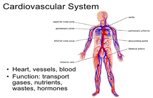
The cardiovascular system is a vital organ system responsible for the circulation of blood throughout the body. It consists of the heart, blood vessels, and blood, and plays a crucial role in transporting gases, nutrients, wastes, and hormones to and from tissues.
Heart: The muscular organ that pumps blood.
Blood Vessels: Channels that carry blood throughout the body.
Blood: The fluid that transports essential substances.
Functions of the Cardiovascular System
Maintaining Blood Pressure (BP): Ensures adequate blood flow to all regions. Normal BP is around 120/80 mmHg and is adjusted based on posture, fluid volume, and exercise.
Thermoregulation: Circulation of warm blood helps dissipate heat and regulate core temperature (optimal: 37°C).
Immune Function: Circulates white blood cells (leukocytes) to reduce infection risk and target infected tissues.
Blood Vessels
Types and Structure
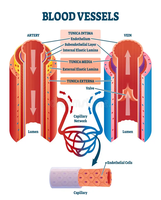
Blood vessels form a closed circuit beginning and ending at the heart. They deliver oxygen and nutrients, remove waste, and are classified into three main types:
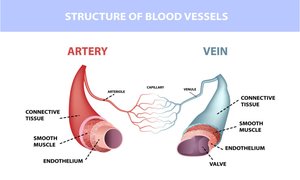
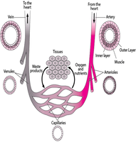
Arteries: Carry blood away from the heart under high pressure.
Veins: Return blood to the heart under low pressure, often containing valves to prevent backflow.
Capillaries: Smallest vessels, connecting arteries and veins, allowing exchange of gases and nutrients.
Arteries and Arterioles
Arteries transport oxygen-rich blood from the heart to the body. Their walls are thick and muscular to withstand high pressure. Arterioles are smaller branches of arteries.
Wall Structure: Three layers: tunica intima, tunica media, tunica externa.
Valves: Not present due to high pressure and unidirectional flow.

Veins and Venules
Veins carry deoxygenated blood back to the heart. Their walls are thinner and contain valves to prevent backflow. Venules are small veins that collect blood from capillaries.
Major Veins: Superior and inferior vena cava.
Pressure: Low, requiring valves for directionality.
Capillaries
Capillaries are thin-walled vessels that facilitate the exchange of oxygen, nutrients, carbon dioxide, and waste between blood and tissues.
Structure: Single layer of endothelial cells.
Function: Connect arteries and veins, allow diffusion.
The Heart
Heart Wall and Pericardial Sack
The heart is encased in the pericardium, a fluid-filled sac that lubricates and protects it. The heart wall consists of three layers:
Endocardium: Inner layer.
Myocardium: Muscular middle layer responsible for contraction.
Pericardium: Protective outer layer.
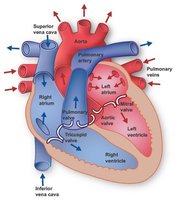
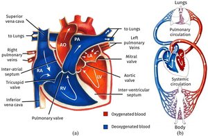
The Four Chambers
The heart is divided into four chambers: two atria (upper) and two ventricles (lower). The right side receives deoxygenated blood and sends it to the lungs; the left side receives oxygenated blood and pumps it to the body.
Right Atrium: Receives blood from the body.
Right Ventricle: Pumps blood to the lungs.
Left Atrium: Receives oxygenated blood from the lungs.
Left Ventricle: Pumps oxygenated blood to the body.
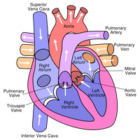
Internal Structures
Interventricular Septum: Separates left and right ventricles.
Trabeculae Carneae: Irregular muscle bands inside ventricles.
Papillary Muscles and Chordae Tendineae: Support AV valves.
Heart Valves and Electrical Conduction
Heart Valves
Valves ensure unidirectional blood flow between chambers and out of the heart:
Atrioventricular (AV) Valves: Tricuspid (right), Mitral/Bicuspid (left).
Semilunar (SL) Valves: Aortic (left ventricle to aorta), Pulmonary (right ventricle to pulmonary artery).
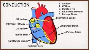
Electrical Conduction System
The heart's electrical system coordinates contraction:
Sinoatrial (SA) Node: Initiates heartbeat.
Atrioventricular (AV) Node: Relays signal to ventricles.
Bundle of His and Purkinje Fibers: Spread signal through ventricles.
The Cardiac Cycle
Phases of the Cardiac Cycle
Each heartbeat consists of contraction (systole) and relaxation (diastole):
Complete Cardiac Diastole: Rest period, myocardium relaxed.
Atrial Systole: Atria contract.
Ventricular Systole: Ventricles contract.
Key Formulas
Cardiac Output:
Blood Pressure:
Blood Flow
Pulmonary and Systemic Circulation
The heart pumps blood into two separate circuits:
Pulmonary Circulation: Right side of heart to lungs and back.
Systemic Circulation: Left side of heart to body and back.
Physiological Measurements
Blood Pressure
Blood pressure is the force exerted by blood on vessel walls, measured in mmHg. It is given as systolic (during contraction) and diastolic (during relaxation) values.
Ideal BP: 90/60 mmHg to 120/80 mmHg
High BP: 140/90 mmHg or higher
Low BP: Below 90/60 mmHg
Heart Rate
Normal adult heart rate is 60–100 bpm at rest. It varies with age, health, and lifestyle. Tachycardia is a fast heart rate; bradycardia is slow.
Respiratory Rate
The cardiovascular and respiratory systems work together to deliver oxygen and remove carbon dioxide. Respiratory rate is measured as breaths per minute, with inspiration and expiration phases.

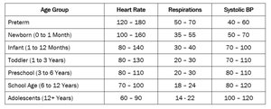
Paediatric Measurements
Normal ranges for heart rate, respiration, and blood pressure vary by age group. The table below summarizes these values:
Age Group | Heart Rate | Respirations | Systolic BP |
|---|---|---|---|
Preterm | 120–150 | 50–70 | 40–60 |
Newborn (0–1 Month) | 100–160 | 35–55 | 50–70 |
Infant (1–12 Months) | 80–140 | 30–40 | 70–100 |
Toddler (1–3 Years) | 80–130 | 20–30 | 80–110 |
Preschool (3–6 Years) | 80–110 | 20–30 | 80–110 |
School Age (6–12 Years) | 70–100 | 18–24 | 80–120 |
Adolescents (12+ Years) | 60–90 | 14–22 | 100–120 |
Temperature Regulation
Normal body temperature ranges from 36.5–37.5°C. The cardiovascular system helps regulate temperature by dilating or constricting blood vessels. Temperature can be measured at various sites: tympanic, axillary, rectal.
Dilation: Releases heat.
Constriction: Retains heat.
References
OpenStax (2023) Anatomy and Physiology.
Tortora, G.J. and Derrickson, B. (2021) Principles of Anatomy and Physiology.
NHS (2023) The heart and circulation.
British Heart Foundation (2022) How the heart works.
Guyton, A.C. and Hall, J.E. (2020) Textbook of Medical Physiology.
