 Back
BackUrinary System: Medical Terminology and Anatomy Study Guide
Study Guide - Smart Notes
 Tailored notes based on your materials, expanded with key definitions, examples, and context.
Urinary System Overview
Introduction to the Urinary System
The urinary system is responsible for filtering metabolic waste from the bloodstream and eliminating it from the body through excretion. It plays a crucial role in maintaining homeostasis by regulating fluid and electrolyte balance, as well as removing toxins.
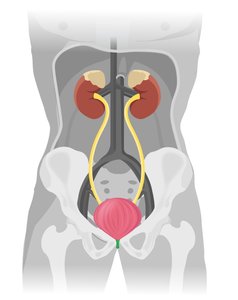
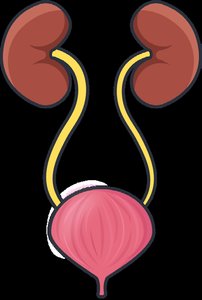
Key Organs: Kidneys, ureters, urinary bladder, and urethra.
Main Function: Filtration of blood, urine production, storage, and excretion.
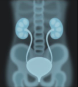
Location: The kidneys are located in the lumbar region above the waist, on either side of the vertebral column.
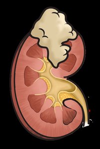
External and Internal Anatomy of the Kidney
External Anatomy
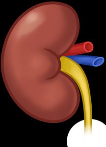
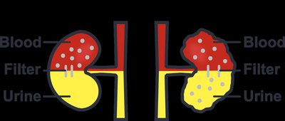
The kidney has a characteristic bean shape with a concave area called the renal hilum on its medial surface. The renal artery enters, and the renal vein and ureter exit through the hilum.
Renal Artery: Supplies blood to the kidney.
Renal Vein: Drains filtered blood from the kidney.
Ureter: Transports urine from the kidney to the bladder.
Internal Anatomy
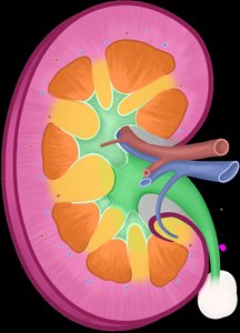
The kidney is divided into three main regions:
Renal Cortex: The outermost region, containing the majority of nephrons.
Renal Medulla: The inner region, organized into triangular structures called renal pyramids. The tip of each pyramid is the renal papilla, which points toward the renal pelvis.
Renal Pelvis: A funnel-shaped cavity that collects urine from the calyces and channels it into the ureter.
Calyx: Cup-shaped tubes that collect urine from the pyramids and empty into the renal pelvis.
Nephron Structure and Function
Nephron: The Functional Unit
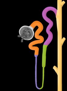
Each kidney contains about one million nephrons, which are responsible for filtering blood and forming urine. The nephron consists of two main parts: the renal corpuscle and the renal tubule.
Renal Corpuscle: Includes the glomerulus (a cluster of capillaries) and the Bowman's (glomerular) capsule (a double-walled structure that encases the glomerulus).
Afferent arteriole: Brings blood to the glomerulus.
Efferent arteriole: Carries blood away from the glomerulus.
Renal Tubule: Reabsorbs needed substances (water, electrolytes) and removes waste. It is divided into:
Proximal convoluted tubule
Nephron loop (Loop of Henle): Descending and ascending limbs
Distal convoluted tubule
Collecting tubule
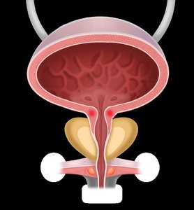
Urinary Bladder Anatomy and Physiology
Structure and Function
The urinary bladder is an elastic, muscular sac located in the anterior portion of the pelvic cavity. It stores urine until excretion. The bladder wall is composed of three layers of smooth muscle and is lined with a mucous membrane containing rugae, which allow expansion.
Internal sphincter: Made of smooth muscle, relaxes involuntarily during urination.
External sphincter: Made of skeletal muscle, under voluntary control.
In males: The prostate gland is located inferior to the bladder.
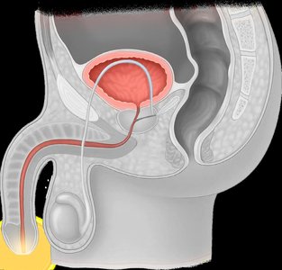
Urethra Anatomy
Male and Female Urethra
The urethra is a tubular structure that carries urine from the bladder to the outside of the body. The external opening is called the urinary meatus.
Female urethra: Shorter and located anterior to the vagina; carries only urine.
Male urethra: Longer, passes through the prostate and penis; carries both urine and semen.

Medical Terminology: Word Building
Combining Forms and Suffixes
Medical terms for the urinary system are constructed using combining forms and suffixes. Understanding these helps in deciphering complex terminology.
Combining Form/Suffix | Definition | Memory Tool |
|---|---|---|
vesic/o | bladder | vessel |
pyel/o | renal pelvis | Kidney Pilot Elf |
azot/o | nitrogenous waste | No No risotto! |
contin/o | to hold in | Contain it! |
noct/o | night | nocturnal |
olig/o | few in number | oligarchy |
spadias/o | tear, slit | spade (split card) |
idi/o | distinctive, unknown | ID |
-tripsy | crushing | Trip to Italy (grape stomping) |


Urinary System: Vocabulary and Pathology
Common Terms and Disorders
Understanding the vocabulary related to urinary system pathology and pharmacology is essential for recognizing clinical conditions and treatments.
Medical Term | Definition | Memory Tool |
|---|---|---|
Enuresis | Involuntary urination (bedwetting) | In Your PJs |
Calculus | Small stone formed from mineral salts | Calculating Abacus |
Stricture | Abnormal narrowing of a urinary passageway | Strictly narrowed structure |
Wilms Tumor | Malignant kidney tumor in children | Welling up Kid-ney |
Thiazide | Diuretic for high BP and edema | Edemic THIgh |
Potassium-Sparing | Diuretic that spares potassium | K stays |
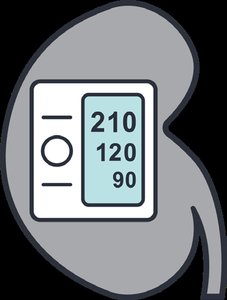
Renin | Enzyme controlling BP via vasoconstriction | Raises Readings |


Urinary Diagnostic and Laboratory Terms
Key Diagnostic Tests
Several laboratory and imaging tests are used to assess kidney function and diagnose urinary system disorders.
Medical Term | Definition | Memory Tool |
|---|---|---|
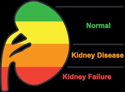
Albumin/Creatinine Ratio (ACR) | Screening test for albumin and creatinine in urine; high ratio indicates kidney disease | Albumin Creates Risks |
Estimated Glomerular Filtration Rate (eGFR) | Measures how effectively kidneys filter blood | Filtration Rate |
Retrograde Pyelography (RP) | X-ray with contrast dye injected against urine flow to visualize kidneys, ureters, bladder | Renally Precise Imaging |
Summary Table: Main Structures and Functions
Structure | Function |
|---|---|
Kidney | Filters blood, forms urine |
Ureter | Transports urine to bladder |
Urinary Bladder | Stores urine |
Urethra | Excretes urine from body |
Key Equations
Estimated Glomerular Filtration Rate (eGFR):
Additional info: The above formula is a simplified version; actual eGFR calculations may use more complex variables and constants depending on the method (e.g., MDRD or CKD-EPI equations).
