 Back
BackUrinary System: Nephron Function, Hormonal Regulation, Kidney Disorders, and Stones
Study Guide - Smart Notes
 Tailored notes based on your materials, expanded with key definitions, examples, and context.
Urinary System
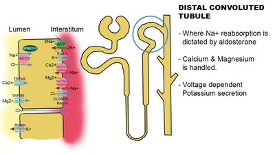
Distal Convoluted Tubule (DCT)
The distal convoluted tubule is a segment of the nephron located after the loop of Henle. It plays a crucial role in the regulation of electrolyte and fluid balance in the body.
Location: Follows the loop of Henle in the nephron structure.
Function: Regulates sodium (Na+) and chloride (Cl−) concentrations in the filtrate through selective reabsorption.
Hormonal Control: Sodium reabsorption is influenced by the hormone aldosterone.
Electrolyte Handling: Responsible for the reabsorption of calcium (Ca2+) and magnesium (Mg2+), and for voltage-dependent potassium (K+) secretion.
Clinical Relevance: Dysfunction can contribute to electrolyte imbalances and hypertension.
Example: Thiazide diuretics act on the DCT to inhibit sodium reabsorption, increasing urine output.
Collecting Duct Function
The collecting duct is the final segment of the nephron and is essential for the regulation of water reabsorption and urine concentration.
Receives Filtrate: Collects filtrate from multiple nephrons.
Water Reabsorption: Major site for water reabsorption, regulated by hormones such as antidiuretic hormone (ADH).
Urine Concentration: Determines the final concentration and volume of urine excreted.
Clinical Relevance: Disorders affecting the collecting duct can lead to diabetes insipidus or inappropriate antidiuretic hormone secretion.
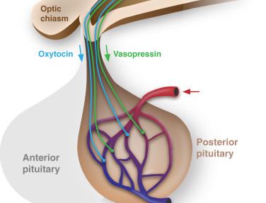
Antidiuretic Hormone (ADH)
ADH, also known as vasopressin, is a hormone that plays a key role in water balance and osmoregulation in the body.
Production: Synthesized in the hypothalamus.
Storage and Release: Stored and released from the posterior pituitary gland.
Function: Released when the body needs to conserve water, such as during dehydration or increased plasma osmolality.
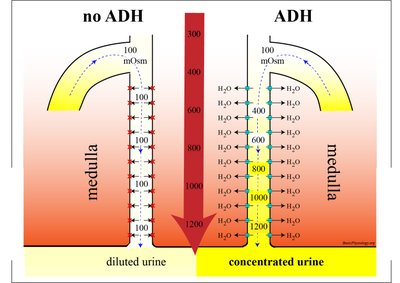
ADH Action on the Collecting Duct
ADH increases the permeability of the collecting duct to water, allowing more water to be reabsorbed back into the bloodstream, thus concentrating the urine.
Mechanism: ADH binds to receptors on the collecting duct cell membranes, triggering the insertion of aquaporin water channels into the membrane.
Result: Increased water reabsorption and reduced urine volume.
Clinical Relevance: Lack of ADH or resistance to its effects leads to excessive urination and dehydration (diabetes insipidus).
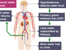
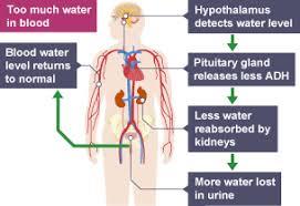
Osmoregulation and ADH
Osmoregulation is the process by which the body maintains the proper balance of water and electrolytes. ADH is a central hormone in this process.
Low Blood Water: Triggers increased ADH release, leading to more water reabsorption and concentrated urine.
High Blood Water: Suppresses ADH release, resulting in less water reabsorption and dilute urine.
Feedback Mechanism: The hypothalamus detects changes in blood osmolality and adjusts ADH secretion accordingly.
Kidney Disorders
Kidney Failure
Kidney failure, or renal failure, occurs when the kidneys lose their ability to filter waste and maintain fluid and electrolyte balance.
Definition: Inability of the kidneys to filter waste and excess fluid effectively, leading to toxin and fluid accumulation.
Types: Acute (sudden onset) and chronic (progressive loss of function).
Treatment: May require dialysis or kidney transplant in severe cases.
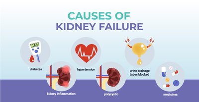
Causes of Kidney Failure
High blood pressure (hypertension)
Diabetes mellitus
Glomerulonephritis (inflammation of the glomerulus)
Genetic kidney conditions (e.g., polycystic kidney disease)
Long-term kidney infections
Symptoms of Kidney Failure
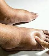
Swelling in ankles or feet (edema) due to fluid retention
Persistent tiredness and fatigue
Shortness of breath (fluid in lungs)
Blood in urine (hematuria)
Infrequent urination
Itchy skin and muscle cramps (waste buildup)
Oedema (Edema)
Oedema is swelling caused by excess fluid trapped in body tissues, often seen in the legs, feet, and ankles. It is a common complication of kidney failure due to impaired fluid regulation.

Treatments for Kidney Failure
Lifestyle changes (healthy diet, exercise, reduced salt intake)
Medications to control blood pressure and cholesterol
Medications to slow kidney damage
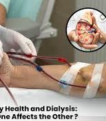
Dialysis to filter blood when kidneys fail
Kidney transplant

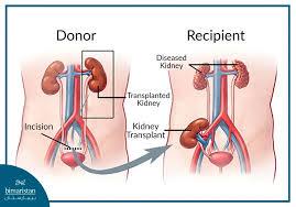
Kidney Transplant
A kidney transplant is a surgical procedure to replace a diseased kidney with a healthy one from a donor. It is considered when dialysis is insufficient or not desired.
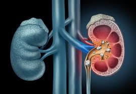
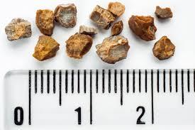
Kidney Stones
Kidney Stones: Definition and Formation
Kidney stones are hard deposits formed from chemicals in urine, such as calcium or uric acid. They develop when these substances crystallize inside the kidney.
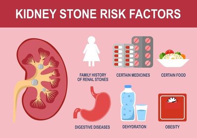
Causes and Risk Factors of Kidney Stones
Inadequate fluid intake (dehydration)
High levels of certain chemicals in urine (e.g., calcium, oxalate, uric acid)
Urinary tract infections
Diet high in salt or protein
Family history of kidney stones
Digestive diseases and obesity
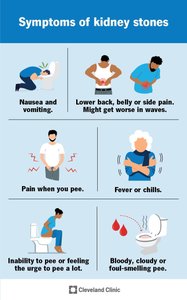
Symptoms of Kidney Stones
Severe side or back pain (renal colic) due to ureter blockage
Pain radiating to the groin as the stone moves
Blood in urine (hematuria) from urinary tract irritation
Nausea or vomiting due to pain
Fever or chills if infection is present
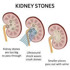
Treatments for Kidney Stones
Pain relief medications (e.g., NSAIDs)
Anti-sickness medications
Increased fluid intake to help pass stones
Alpha-blocker medications to relax ureter muscles
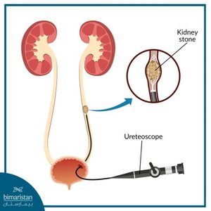
Surgical removal or procedures for large stones
Lithotripsy (shock wave therapy to break up stones)
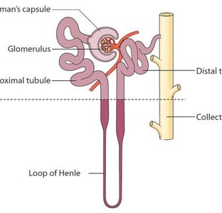
Additional info: The nephron is the functional unit of the kidney, and its segments (including the DCT and collecting duct) are essential for urine formation and homeostasis. Disorders of these structures can lead to significant clinical symptoms and require targeted treatments.
