 Back
BackBiological Membranes and Transport: Mini-Textbook Study Guide
Study Guide - Smart Notes
 Tailored notes based on your materials, expanded with key definitions, examples, and context.
Biological Psychology
Biological Membranes and Transport
Biological membranes are fundamental to cellular function, acting as selective barriers that regulate the movement of molecules into and out of cells. Understanding membrane transport is essential for grasping physiological processes, including neural signaling, metabolism, and homeostasis.
Structure and Selectivity of Biological Membranes
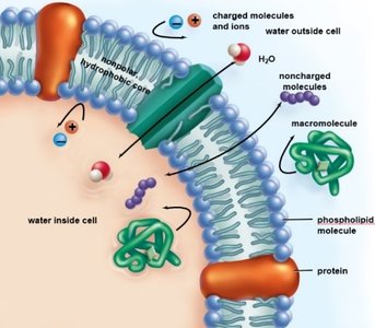
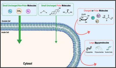
Phospholipid Bilayer: The cell membrane is primarily composed of a phospholipid bilayer with embedded proteins, creating a nonpolar, hydrophobic core that restricts passage of certain molecules.
Selective Permeability: Membranes are selectively permeable, allowing some molecules to cross freely while restricting others based on size, charge, and polarity.
Key Components: Phospholipids, proteins, and cholesterol contribute to membrane structure and function.
Example: Small, nonpolar molecules (e.g., O2, CO2) diffuse easily, while large or charged molecules require facilitation.
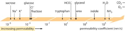
Diffusion and Permeability
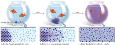
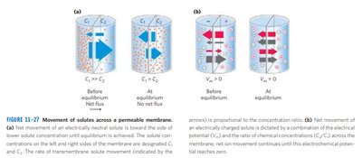
Diffusion: Molecules move from areas of high concentration to low concentration, down their concentration gradient.
Freely Diffusing Molecules: Uncharged, nonpolar, hydrophobic molecules can cross the membrane without facilitation.
Restricted Molecules: Large, charged, or hydrophilic molecules cannot cross freely and require transport proteins.
Permeability Coefficient: Quantifies how easily a molecule crosses the membrane; higher values indicate greater permeability.
Example: O2 and CO2 have high permeability, while Na+ and K+ have low permeability.
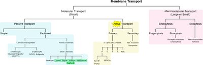
Types of Membrane Transport
Passive Transport: Does not require energy; includes simple diffusion and facilitated diffusion.
Active Transport: Requires energy (usually ATP) to move molecules against their concentration gradient.
Transport Proteins: Include channels, carriers, and pumps, each with specific mechanisms and selectivity.
Example: Glucose transporters (GLUT) facilitate passive transport, while Na+/K+ ATPase performs active transport.
Classes of Membrane Transport Proteins
Uniporters: Transport one molecule at a time in one direction.
Symporters: Cotransport two or more molecules in the same direction.
Antiporters: Cotransport two or more molecules in opposite directions.
Example: The Cl–/HCO3– antiporter exchanges chloride and bicarbonate ions across erythrocyte membranes.
Passive Transport: Simple vs. Facilitated Diffusion
Simple Diffusion: Direct movement through the membrane without assistance.
Facilitated Diffusion: Requires a membrane protein (channel or carrier) but does not require energy.
Both: Move molecules down their concentration gradient.
Example: GLUT1 transporter in erythrocytes facilitates glucose uptake.
Kinetics of Passive Transport
Simple Diffusion: Rate increases linearly with concentration gradient.
Facilitated Diffusion: Rate increases rapidly but plateaus as transport proteins become saturated, forming a Michaelis-Menten curve.
Ktr: Substrate concentration at which the transport protein is half-saturated (analogous to Km in enzyme kinetics).
Facilitated Diffusion: Carrier and Channel Proteins
Carrier/Transporter: Undergoes conformational changes to move solutes across the membrane.
Porin/Channel: Forms a membrane-spanning pore for rapid transport of ions or water.
Aquaporins: Specialized channels for water transport.
Example: Movement of water across membranes is facilitated by aquaporins.
Erythrocyte Transport Models
GLUT1 Uniporter: Facilitates glucose transport into erythrocytes, maintaining low intracellular glucose due to metabolism.
Cl–/HCO3– Antiporter: Mediates chloride shift, crucial for CO2 transport and blood pH regulation.
Chloride Shift: Exchange of Cl– and HCO3– near tissues and lungs, maintaining ionic balance and CO2 transport.
Membrane Transport of Ions
Electrochemical Gradient: Combination of chemical concentration and electrical potential gradients determines ion movement.
Transmembrane Potential (ΔΨ): Difference in electrical charge across the membrane, usually negative inside cells.
Ion Channels: Selectively transport ions; types include leak, ligand-gated, signal-gated, voltage-gated, and mechanically-gated channels.
Example: Voltage-gated potassium channels are essential for action potentials.
Active Membrane Transport
Primary Active Transport: Directly uses energy (ATP hydrolysis) to move molecules against their gradient.
Secondary Active Transport: Uses electrochemical gradients established by primary active transport to drive movement of other molecules.
Types of ATPases: P-type, V-type, F-type, A-type, and ABC transporters, each with specific substrates and mechanisms.
Type | Function | Example |
|---|---|---|
P-type ATPase | Transports cations, reversibly phosphorylated | Na+/K+ pump |
V-type ATPase | Transports H+ to acidify vesicles | Lysosomes |
F-type ATPase | Transports H+ to produce ATP | ATP synthase |
A-type ATPase | Transports anions | Archaeal membranes |
ABC Transporter | Pumps various solutes, multidrug resistance | P-glycoprotein |
Sodium-Potassium Ion Pump (Na+/K+ ATPase)
Maintains: Low intracellular Na+ and high K+, establishing electrical and chemical gradients.
Mechanism: Pumps 3 Na+ out and 2 K+ in per ATP hydrolyzed, creating a negative membrane potential.
Example: Essential for nerve impulse transmission and muscle contraction.
SERCA: Calcium Ion Pump
SERCA: Sarcoplasmic/Endoplasmic Reticulum Ca2+ ATPase, a P-type ATPase.
Function: Pumps Ca2+ into SR/ER, keeping cytoplasmic Ca2+ low.
Role: Regulates muscle contraction and relaxation.
ABC Transporters and Multidrug Resistance
ABC Transporters: ATP-Binding Cassette proteins with two transmembrane domains and two nucleotide-binding domains.
Function: Pump substances against their gradient, including drugs and toxins.
Example: P-glycoprotein confers resistance to anticancer drugs in tumor cells.
Secondary Active Transport: Na+-Glucose Symporter
Mechanism: Uses Na+ gradient (established by Na+/K+ ATPase) to import glucose against its gradient.
Location: Intestinal epithelial cells.
Example: Glucose is transported into cells via symport with Na+, then exported to blood via GLUT2.
Endocytosis and Exocytosis
Endocytosis: Uptake of large molecules via vesicle formation; includes phagocytosis, pinocytosis, and receptor-mediated endocytosis.
Exocytosis: Release of molecules from cells via vesicle fusion with the membrane; essential for neurotransmitter and hormone secretion.
Fusion Proteins: SNARE proteins mediate vesicle fusion during exocytosis.
Thermodynamics of Membrane Diffusion
Gibbs Free Energy (ΔG): Determines spontaneity of transport.
Uncharged Molecules:
Charged Ions:
Variables: R = gas constant, T = temperature (K), z = ion charge, F = Faraday constant, ΔΨ = membrane potential.
Example: Movement of Na+ into a cell is favored when ΔG is negative.
Summary Table: Membrane Transport Types
Transport Type | Energy Requirement | Direction | Protein Involvement |
|---|---|---|---|
Simple Diffusion | No | Down gradient | No |
Facilitated Diffusion | No | Down gradient | Yes |
Primary Active Transport | Yes (ATP) | Against gradient | Yes |
Secondary Active Transport | Indirect (gradient) | Against gradient | Yes |
Endocytosis/Exocytosis | Yes | Bulk transport | Yes (fusion proteins) |
Additional info: These notes expand on the original content by providing definitions, examples, and academic context for each transport mechanism, including relevant equations and tables for clarity.
