 Back
BackBiological Psychology: Neurons, Neurotransmitters, and the Brain
Study Guide - Smart Notes
 Tailored notes based on your materials, expanded with key definitions, examples, and context.
Biological Psychology
Facilitated Communication (FC) and Scientific Skepticism
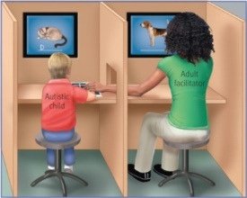
Facilitated Communication (FC) is a controversial technique once used to assist individuals with severe communication impairments, such as autism, in expressing themselves. In FC, a facilitator supports the hand or arm of the individual as they type or point to letters or pictures. However, scientific investigations have raised significant doubts about its validity.
Definition of FC: A method where a facilitator physically supports a nonverbal person's hand, allegedly helping them communicate.
Scientific Skepticism: Many researchers were skeptical because evidence suggested facilitators, not the clients, were the true source of the messages.
Testing FC: Controlled experiments separated the facilitator and client, showing that correct answers only appeared when the facilitator knew the answer, not the client.
Ethical Concerns: Belief in FC can lead to serious harm, such as false accusations, wrongful imprisonment, and inappropriate medical or legal decisions.
Critical Thinking: The "not me fallacy" is the belief that one is immune to errors in thinking that affect others, which can perpetuate belief in pseudoscientific methods like FC.

Additional info: FC is a classic example of why scientific thinking and skepticism are essential in psychology, as it demonstrates the dangers of untested or disproven interventions.
Neurons: Structure and Function
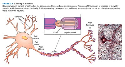
Basic Anatomy of a Neuron
Neurons are specialized cells that transmit information throughout the central nervous system (CNS). They communicate via electrical and chemical signals, forming the basis of all thoughts, behaviors, and bodily functions.
Cell Body (Soma): Contains the nucleus and is responsible for the cell's metabolic activities.
Dendrites: Branch-like structures that receive messages from other neurons and transmit them toward the cell body.
Axon: A long, slender projection that carries electrical impulses away from the cell body to other neurons or muscles.
Myelin Sheath: Fatty insulation that covers some axons, speeding up neural transmission. Damage to myelin (e.g., in multiple sclerosis) impairs signal conduction.
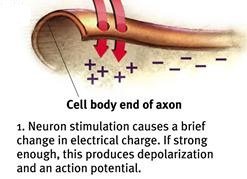
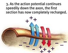
Action Potential: The brief electrical charge that travels down the axon when a neuron fires. This process is "all or none"—the neuron either fires completely or not at all.
Resting Potential: The state of a neuron when not firing, with a negative charge inside the axon relative to the outside.
Action Potential and Neural Transmission
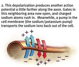
The action potential is generated by the movement of ions across the neuron's membrane, leading to a rapid change in electrical charge that propagates along the axon.
Depolarization: When stimulated, sodium ions rush into the axon, making the inside more positive and triggering the action potential.
Repolarization: Potassium ions exit the cell, restoring the negative charge inside.
Sodium-Potassium Pump: Actively restores the original ion balance after the action potential passes.
Synaptic Transmission
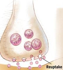
The Synapse and Neurotransmitter Release
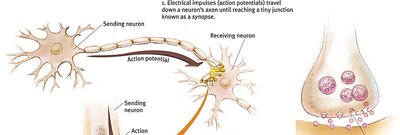
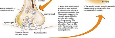
Neurons communicate at synapses, where the axon terminal of one neuron meets the dendrite of another. Chemical messengers called neurotransmitters are released into the synaptic cleft and bind to receptor sites on the receiving neuron.
Synaptic Vesicles: Store neurotransmitters in the axon terminal.
Neurotransmitter Release: Triggered by the arrival of an action potential at the axon terminal.
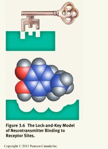
Receptor Sites: Specific locations on the receiving neuron where neurotransmitters bind, following a "lock-and-key" model.
Reuptake: The process by which excess neurotransmitters are reabsorbed by the sending neuron.
Major Neurotransmitters and Their Functions
Types and Effects of Neurotransmitters
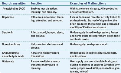
Neurotransmitters are chemicals that transmit signals across synapses. Each has specific functions and is linked to various psychological and physiological processes.
Neurotransmitter | Function | Examples of Malfunctions |
|---|---|---|
Acetylcholine (ACh) | Enables muscle action, learning, memory | With Alzheimer's disease, ACh-producing neurons deteriorate |
Dopamine | Influences movement, learning, attention, emotion | Excess receptor activity linked to schizophrenia; undersupply leads to Parkinson's disease |
Serotonin | Affects mood, hunger, sleep, arousal | Undersupply linked to depression; antidepressants raise serotonin levels |
Norepinephrine | Helps control alertness and arousal | Undersupply can depress mood |
GABA | Major inhibitory neurotransmitter | Undersupply linked to seizures, tremors, insomnia |
Glutamate | Major excitatory neurotransmitter; involved in memory | Oversupply can overstimulate brain, causing migraines or seizures |
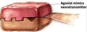
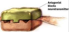
Agonists and Antagonists
Drugs and other chemicals can affect neurotransmitter activity by acting as agonists (mimicking neurotransmitters) or antagonists (blocking neurotransmitters).
Agonists: Imitate neurotransmitters and activate receptors (e.g., morphine mimics endorphins).
Antagonists: Block neurotransmitter action by occupying receptor sites (e.g., naloxone blocks opiate effects).
The Brain: Structure and Function
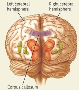
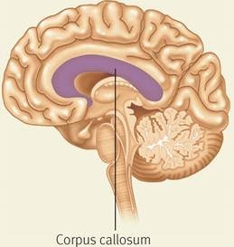
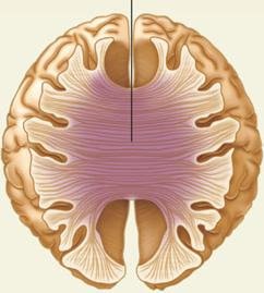
Cerebral Hemispheres and Corpus Callosum
The cerebrum is divided into two hemispheres, connected by the corpus callosum. Each hemisphere specializes in certain functions but communicates with the other for integrated processing.
Left Hemisphere: Language, logic, analytical tasks.
Right Hemisphere: Spatial abilities, face recognition, creative tasks.
Corpus Callosum: Thick band of fibers connecting the hemispheres, allowing information transfer.

Lobes of the Cerebral Cortex
The cerebral cortex is divided into four lobes, each with specialized functions:
Frontal Lobe: Movement, problem-solving, decision-making, planning, language.
Parietal Lobe: Touch, spatial ability, attention.
Occipital Lobe: Vision, shape and form recognition.
Temporal Lobe: Hearing, memory, language, complex vision (e.g., face recognition).
Brain Mapping Techniques
Modern neuroscience uses various techniques to study brain structure and function:
EEG (Electroencephalogram): Measures electrical activity in the brain; good for timing, not location.
MRI (Magnetic Resonance Imaging): Provides detailed images of brain structure.
fMRI (Functional MRI): Measures brain activity by detecting changes in blood oxygenation.
PET (Positron Emission Tomography): Tracks glucose consumption to show active brain areas.
TMS (Transcranial Magnetic Stimulation): Uses magnetic fields to stimulate or disrupt brain regions.
Key Brain Structures and Their Functions
Brainstem: Includes the medulla (controls vital functions), pons (sleep, facial movement), and reticular formation (arousal, attention).
Limbic System: Includes the amygdala (emotion, fear), hippocampus (memory formation), thalamus (sensory relay), and hypothalamus (regulation of hunger, thirst, temperature, sexual behavior).
Cerebellum: Coordinates movement, balance, and procedural memory.
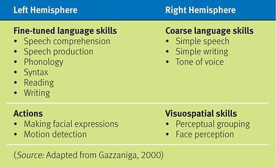
Split-Brain Research and Hemispheric Specialization
Split-brain studies (e.g., after corpus callosum surgery) reveal that each hemisphere has specialized functions, but both are necessary for integrated behavior. The myth that people are strictly "right-brained" or "left-brained" is not supported by evidence.
Left Hemisphere | Right Hemisphere |
|---|---|
|
|
Review Questions and Applications
Understanding the structure and function of the nervous system is essential for explaining behavior, diagnosing disorders, and developing treatments. The review questions provided test knowledge of neural conduction, neurotransmitter function, brain anatomy, and the consequences of brain injury.
