 Back
BackBiological Psychology: Structure and Function of the Nervous System
Study Guide - Smart Notes
 Tailored notes based on your materials, expanded with key definitions, examples, and context.
Biological Psychology
Introduction to Biological Psychology
Biological psychology explores the relationship between the nervous system and behaviour, focusing on how brain structure and function influence psychological processes. Researchers in this field are known as biological psychologists or neuroscientists.
Key Point: Biological psychology bridges neuroscience and psychology, examining how neural mechanisms underpin behaviour.
Example: Studying how neurotransmitter imbalances contribute to mental disorders.
Brain Mapping Methods
Historical and Modern Techniques
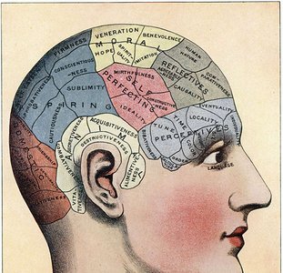
Brain mapping methods are used to study the structure and function of the brain. Early methods like phrenology, which linked skull shape to cognitive abilities, have been discredited. Modern techniques include neuropsychological tests, brain lesion studies, and advanced imaging technologies.
Phrenology: Early, now discredited, method that attempted to link skull shape to mental faculties.
Neuropsychology: Uses tests to assess cognitive function in individuals with brain damage, considering language and cultural influences.
Animal Studies: Controlled brain lesions in animals to study behaviour.
Electroencephalograph (EEG)
EEG measures electrical activity in the brain using electrodes placed on the scalp. It is excellent for studying changes in brain activity over milliseconds but less effective for pinpointing small regions. 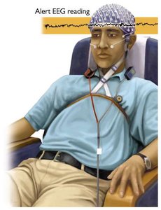
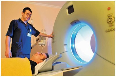
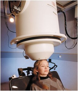
Neuroimaging Techniques
Neuroimaging allows visualization of brain structure and function.
Computed Tomography (CT): Uses X-rays to create 3D images of the brain.
Magnetic Resonance Imaging (MRI): Uses magnetic fields to visualize brain structure.
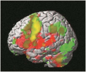
Functional MRI (fMRI): Visualizes brain activity by detecting changes in blood flow.
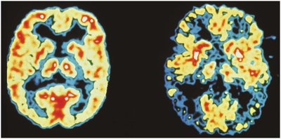
Positron Emission Tomography (PET): Measures glucose consumption to map neural activity.
Magnetoencephalography (MEG): Measures tiny magnetic fields generated by the brain.
Brain Stimulation Techniques
Deep Brain Stimulation (DBS): Uses implanted electrodes to modify brain function, potentially treating disorders like Parkinson’s disease and depression.
Transcranial Magnetic Stimulation (TMS): Applies magnetic fields to the skull to enhance or interrupt brain function.
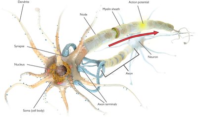
Neurons: The Brain’s Communicators
Structure and Function of Neurons
Neurons are specialized cells for communication, with approximately 86 billion neurons and 160 trillion synaptic connections in the human brain. 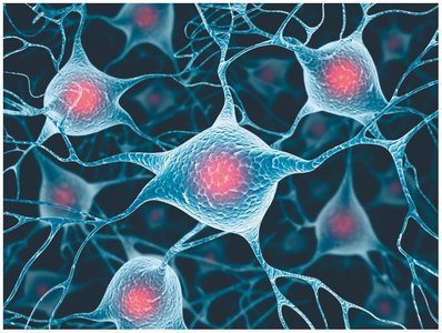
Cell Body (Soma): Builds new cell components.
Dendrites: Receive information from other neurons.
Axons: Transmit information to other neurons.
Axon Terminal: Contains synaptic vesicles filled with neurotransmitters.
Synapse: The gap between neurons where neurotransmitters are released.
Synaptic Transmission
Synaptic transmission is the process by which neurotransmitters are released from one neuron and bind to receptors on another, enabling communication. 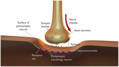
Electrical Responses of Neurons
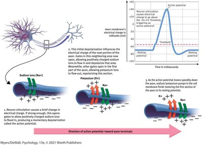
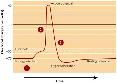
Action Potential
The action potential is an abrupt wave of electrical discharge that travels down the axon, triggering neurotransmitter release. It is an all-or-none response, originating near the cell body and moving to the axon terminal.
Resting Potential: The membrane potential when the neuron is not stimulated, typically around -60 mV.
Threshold of Excitation: The level of stimulation required to trigger an action potential.
Refractory Period: Brief period after firing when the neuron cannot fire again.
Glial Cells
Types and Functions
Glial cells support neurons and are equally numerous in the brain.
Astrocytes: Star-shaped, most abundant, increase reliability of neuronal transmission, found in the blood-brain barrier.
Oligodendrocytes: Promote new connections and produce the myelin sheath around axons.
Neurotransmission
Neurotransmitters and Communication
Communication within neurons is electrical, while communication between neurons is chemical. Neurotransmitters bind to receptor sites on the next neuron, following a lock-and-key mechanism. 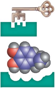
Reuptake: Process where neurotransmitters are reabsorbed into the axon terminal.
Excitatory vs. Inhibitory: Some neurotransmitters excite, others inhibit neural activity.
Types of Neurotransmitters
Glutamate: Excitatory, involved in learning and memory; toxic in high doses.
GABA: Inhibitory, dampens neural activity.
Acetylcholine: Influences arousal, attention, sleep, memory, and muscle movement.
Monoamines: Includes norepinephrine (arousal, mood), dopamine (motor function, reward), serotonin (mood, sleep).
Anandamides: Influence eating, motivation, memory, sleep; bind to same receptors as THC.
Neuropeptides: Short amino acid chains; endorphins relieve pain, others regulate hunger and memory.
Psychoactive Drugs
Effects on Neurotransmitter Systems
Psychoactive drugs interact with neurotransmitter systems, affecting mood, arousal, and behaviour.
Agonists: Increase activity of neurotransmitter systems (e.g., opiates mimic endorphins).
Antagonists: Decrease activity (e.g., dopamine blockers for schizophrenia).
Neural Plasticity
Development and Learning
Plasticity refers to the nervous system’s ability to change. Neurons change through growth, synaptogenesis, pruning, and myelination. Learning can potentiate synapses.
Adult Plasticity: Decreases with age; recovery from injury is limited.
Stem Cells: Can become any cell, including neurons; potential for neurogenesis.
Brain-Behaviour Network
Central and Peripheral Nervous Systems
Sensory information enters and decisions exit the central nervous system (CNS). The peripheral nervous system (PNS) consists of nerves outside the CNS. 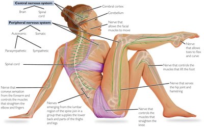
Central Nervous System
Protection and Structure
The CNS is protected by meninges and cerebrospinal fluid-filled ventricles. 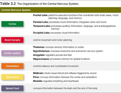
Organization of the CNS
The brain is divided into systems based on location and function. 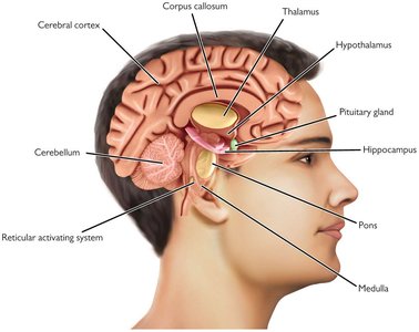
Cerebral Cortex
Lobes and Functions
The cerebral cortex is the largest part of the forebrain, divided into four lobes: frontal, parietal, temporal, and occipital, each with distinct functions. 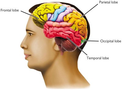
Hemispheres and Corpus Callosum
The forebrain consists of two hemispheres connected by the corpus callosum, enabling communication. 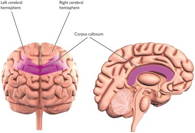
Lateralization
Some cognitive functions are more dominant in one hemisphere.
Right Hemisphere | Left Hemisphere |
|---|---|
Coarse language skills, tone of voice, visuospatial skills, perceptual grouping, face perception | Fine-tuned language skills, speech comprehension and production, reading, writing, making facial expressions, motion detection |
Localization of Function
Brain Regions and Complex Tasks
Many brain areas are associated with specific functions, but complex tasks require coordination across multiple regions.
Split Brain Surgery
Severing the corpus callosum can reduce epileptic seizures, illustrating the importance of interhemispheric communication. 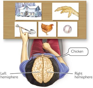
Frontal Lobes
Functions and Mapping
The frontal lobes assist in motor function, language, memory, and executive functioning. Broca’s area is involved in speech production; the motor cortex maps the body. 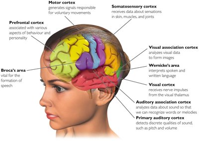
Phineas Gage Case
Phineas Gage’s injury to his prefrontal cortex led to dramatic behavioural changes, illustrating the role of this region in personality and executive function. 
Parietal Lobe
Somatosensory Cortex
The parietal lobe contains the somatosensory cortex, sensitive to pressure, pain, and temperature, and communicates with the motor cortex.
Temporal Lobe
Auditory and Language Processing
The temporal lobe plays a role in hearing, language comprehension (Wernicke’s area), and memory. Lesions can cause neglect and other deficits.
Occipital Lobe
Vision
The occipital lobe is specialized for vision and located at the back of the brain.
Cortical Hierarchies
Sensory Processing
Sensory information first enters the primary sensory cortex, then moves to association cortex for more complex processing.
Basal Ganglia
Movement and Reward
The basal ganglia are forebrain structures that help control movement and reinforcement. Damage can contribute to Parkinson’s disease.
Limbic System
Emotion, Motivation, and Memory
The limbic system is the emotional centre of the brain, involved in smell, motivation, and memory.
Hypothalamus: Regulates internal bodily states and controls the pituitary gland.
Thalamus: Relays sensory information to the cortex.
Amygdala: Key role in fear, excitement, and arousal.
Hippocampus: Spatial memory; damage causes inability to form new memories.
Brain Stem
Basic Functions and Structure
The brain stem connects the cerebral cortex and spinal cord, performing basic bodily functions and serving as a relay station.
Cerebellum: Balance and movement coordination.
Pons: Connects cortex to cerebellum, triggers dreams.
Medulla: Regulates breathing and heartbeat.
Spinal Cord
Signal Transmission and Reflexes
The spinal cord conveys signals between the brain and body, with sensory nerves carrying information to the brain and motor nerves carrying information to the body. Interneurons allow reflexes.
Peripheral Nervous System
Somatic and Autonomic Branches
The PNS is divided into the somatic nervous system (controls voluntary movement) and the autonomic nervous system (controls involuntary actions).
Sympathetic Division: Engaged during crisis (fight or flight).
Parasympathetic Division: Controls rest and digestion.
Endocrine System
Hormones and Behaviour
The endocrine system consists of glands and hormones that regulate bodily functions and emotions.
Pituitary Gland: Controlled by the hypothalamus, releases hormones influencing growth and other functions, including oxytocin.
Adrenal Glands: Release adrenaline and cortisol during emotional arousal.
Sexual Reproductive Glands: Testes and ovaries produce testosterone and estrogen.
Genetics and Behaviour
Nature vs. Nurture
Genes are carried on chromosomes; humans have 46 chromosomes.
Genotype: Set of genes an individual has.
Phenotype: Observable traits.
Dominant/Recessive Genes: Dominant genes mask others; recessive genes are masked.
Epigenetics: Gene expression can be modified by life experiences.
Behavioural Adaptation and Evolution
Adaptation and Fitness
Organisms develop adaptations to better survive and reproduce.
Fitness: Ability to survive and reproduce at higher rates.
Natural Selection: Adaptations become more common in the population.
Behavioural Genetics
Heritability and Research Designs
Behavioural genetics studies the impact of genes and environment on psychological traits, estimating heritability.
Family Studies: Examine trait similarities among relatives.
Twin Studies: Compare identical and fraternal twins.
Adoption Studies: Compare adopted children to biological and adoptive parents.
Summary Table: Organization of the Central Nervous System
System | Main Function |
|---|---|
Cortex | Frontal: executive functions; Parietal: touch, vision; Temporal: auditory, language, memory; Occipital: visual information |
Basal Ganglia | Movement and motor planning |
Limbic System | Thalamus: sensory relay; Hypothalamus: endocrine/autonomic control; Amygdala: arousal/fear; Hippocampus: spatial memory |
Cerebellum | Balance and movement coordination |
Brain Stem | Midbrain: movement/reflexes; Pons: cortex-cerebellum relay; Medulla: breathing/heartbeat |
Spinal Cord | Signal transmission between brain and body |

