 Back
Biological Psychology: Structure and Function of the Brain and Neurons
Study Guide - Smart Notes
 Tailored notes based on your materials, expanded with key definitions, examples, and context.
Biological Psychology
Introduction to Neuroscience
Biological psychology explores the relationship between the brain, nervous system, and behavior. Understanding the structure and function of the brain and neurons is fundamental to this field.
Brain Facts: The human brain weighs about 3 pounds, consists of 65% fat, and uses 20% of the body's energy. It contains approximately 86 billion neurons, and information can travel at speeds up to 268 mph.
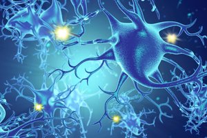
Neurons: Specialized cells that transmit information throughout the nervous system.
Glia: Support cells in the nervous system, including astroglia, oligodendroglia, and microglia.
Phineas Gage: A Case Study in Brain Injury
The case of Phineas Gage is a landmark in neuroscience, demonstrating how brain injury can affect personality and behavior. Gage survived a severe accident in which a tamping iron passed through his skull, damaging his frontal lobe.
Frontal Lobe Damage: Led to significant changes in Gage's personality and social behavior.
Importance: Provided early evidence that specific brain regions are linked to distinct psychological functions.


Structure and Function of the Brain
Major Divisions of the Brain
The brain is divided into three main regions: hindbrain, midbrain, and forebrain. Each region contains specialized structures responsible for different functions.
Hindbrain: Includes the medulla, pons, cerebellum, and reticular formation. Responsible for basic life functions such as heartbeat, breathing, motor coordination, and sleep.
Midbrain: Contains the substantia nigra, important for movement fluidity and inhibition.
Forebrain: Includes the thalamus, hypothalamus, pituitary gland, limbic system, basal ganglia, and cerebral cortex. Responsible for complex functions like motivation, emotion, memory, and cognition.
Forebrain Structures and Functions
Limbic System: Regulates motivation, emotion, learning, and memory. Includes the amygdala (emotion processing) and hippocampus (learning and memory).
Thalamus: Acts as a relay station for sensory information.
Hypothalamus: Controls motivation, basic drives, and the endocrine system.
Pituitary Gland: Regulates hormones.
Basal Ganglia: Involved in voluntary movement and cognitive flexibility. Includes the nucleus accumbens (motivation and reward learning).
Cerebral Cortex: Responsible for consciousness, language, and thought. Divided into lobes: occipital (vision), temporal (auditory/language), parietal (sensory integration), frontal (higher thinking), and prefrontal (memory, morality, planning).
Language Areas of the Brain
Specific regions of the cerebral cortex are involved in language production and comprehension.
Broca's Area: Damage leads to Broca's aphasia, characterized by difficulty producing coherent speech (agrammatism).
Wernicke's Area: Damage results in Wernicke's aphasia, affecting language comprehension and communication.
Corpus Callosum
The corpus callosum connects the right and left hemispheres of the brain. Damage can cause split-brain syndrome, where hemispheres act independently.
Neurons and Neural Communication
Types of Neurons
Neurons are classified based on their function:
Sensory Neurons (Afferent): Carry sensory information to the central nervous system (CNS).
Motor Neurons (Efferent): Transmit motor commands from the CNS to muscles.
Interneurons: Connect sensory and motor neurons within the CNS.
Glial Cells
Astroglia: Create the blood-brain barrier, influence neuron communication, and aid in healing.
Oligodendroglia: Provide myelin to speed up neural transmission.
Microglia: Clean up dead cells and prevent infection.
Action Potential and Neural Transmission
Neurons communicate via electrical impulses called action potentials.
Resting Potential: The neuron is negatively charged inside relative to outside, typically at -70 millivolts.
Action Potential: When a neuron fires, ion channels open, and the inside becomes more positive (+30 millivolts). This triggers neurotransmitter release.
Nodes of Ranvier: Gaps in myelin sheath where action potentials jump, speeding transmission.
Equation:
Where is the change in membrane potential during an action potential.
Synaptic Transmission
Neurons communicate across synapses using neurotransmitters.
Neurotransmitters: Chemicals released into the synapse, binding to receptors on the postsynaptic neuron.
Postsynaptic Potentials: Electrical events in the postsynaptic neuron. Excitatory (EPSPs) increase likelihood of firing; inhibitory (IPSPs) decrease it.
Neuroimaging Techniques
Modern neuroscience uses imaging techniques to study brain structure and activity in awake humans.
MRI (Magnetic Resonance Imaging): Provides detailed images of brain structure.
fMRI (Functional MRI): Measures brain activity by detecting changes in blood flow.
CAT/CT (Computed Axial Tomography): Uses X-rays to create cross-sectional images.
PET (Positron Emission Tomography): Tracks metabolic activity.
TMS (Transcranial Magnetic Stimulation): Stimulates brain regions.
DTI (Diffusion Tensor Imaging): Maps neural pathways.
Comparison of Brain Imaging Techniques
Technique | Main Purpose | Type |
|---|---|---|
MRI | Structural imaging | Non-invasive |
fMRI | Functional imaging | Non-invasive |
CAT/CT | Structural imaging | Non-invasive |
PET | Functional imaging | Invasive (radioactive tracer) |
TMS | Brain stimulation | Non-invasive |
DTI | Mapping neural pathways | Non-invasive |
Additional info: Expanded explanations and context were added to ensure completeness and clarity for introductory psychology students.
