 Back
BackBrain Control of Movement: Hierarchical and Neural Pathways in Motor Function
Study Guide - Smart Notes
 Tailored notes based on your materials, expanded with key definitions, examples, and context.
Brain Control of Movement
Hierarchy of Motor Control
The brain exerts control over movement through a hierarchical system, influencing the motor activity of the spinal cord and initiating voluntary movements. This hierarchy consists of three levels:
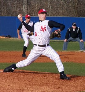
Highest level: Strategy – Involves the neocortex and basal ganglia, responsible for planning the overall goal of movement (e.g., deciding to pitch a ball).
Middle level: Tactics – Includes the motor cortex and cerebellum, responsible for selecting the sequence of muscle contractions (e.g., choosing a particular pitch).
Lowest level: Execution – Comprises the brain stem and spinal cord, responsible for carrying out the movement (e.g., throwing the ball).
Sensorimotor system: Sensory information is used at all levels to refine and guide motor actions.
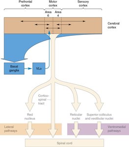
Descending Spinal Tracts
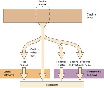
The brain communicates with the spinal cord via descending spinal tracts, which are divided into two major pathways:
Lateral pathways: Control voluntary movement of distal musculature (arms and fingers), under direct cortical control.
Ventromedial pathways: Control posture and locomotion, under brain stem control.
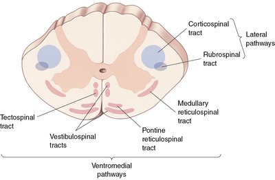
Lateral Pathways
Corticospinal and Rubrospinal Tracts
The lateral pathways are essential for voluntary movement, especially fine motor control of the arms and fingers. They consist of:
Corticospinal tract: The most important component, originating mostly in the motor cortex. It crosses over in the medullary pyramid, so the right motor cortex controls the left side of the body.
Rubrospinal tract: Originates in the red nucleus of the midbrain; its function is reduced in humans.
Effects of lesions: Damage to the corticospinal tract results in deficits in fractionated movement of arms and hands, but posture remains normal. Recovery is possible if the rubrospinal tract is intact.
Clinical relevance: Strokes often damage the motor cortex or corticospinal tract, causing paralysis on the contralateral side, with some recovery over time.
Ventromedial Pathways
Vestibulospinal, Tectospinal, and Reticulospinal Tracts
Ventromedial pathways are crucial for maintaining posture, balance, and locomotion. They use sensory information from the inner ear, visual environment, and proprioceptive inputs.
Vestibulospinal tract: Originates in vestibular nuclei; controls limb and head position for balance.
Tectospinal tract: Originates in the superior colliculus; mediates reflex postural movements of the head in response to visual and auditory stimuli.
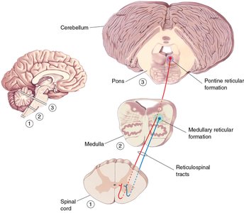
Pontine (medial) reticulospinal tract: Enhances antigravity reflexes by exciting extensor muscles.
Medullary (lateral) reticulospinal tract: Inhibits extensor muscles, liberating them from reflex control.
Motor Planning by the Cerebral Cortex
Motor Cortex Areas and Somatotopic Organization
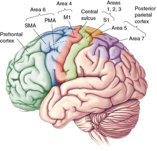
The neocortex, particularly areas 4 (M1) and 6 (SMA+PMA) of the frontal lobe, is involved in voluntary movement control. Area 4 (M1) is the primary motor cortex, where stimulation elicits muscle twitches in specific body regions. Area 6 is a higher motor area, where stimulation elicits complex movements.
PMA (Premotor Area): Lateral region of area 6, involved in planning movement.
SMA (Supplementary Motor Area): Medial region of area 6, involved in coordinating movements.

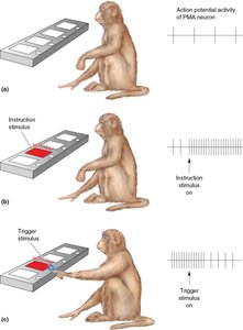
Neuronal Correlates of Motor Planning
Neurons in the PMA fire before movement is performed, indicating their role in motor planning. In experiments with monkeys, PMA neurons begin firing during the "set" phase and continue until movement is executed.
Ready: Parietal and frontal lobes are engaged.
Set: PMA neuron starts firing.
Go: PMA neuron continues firing until movement is made.
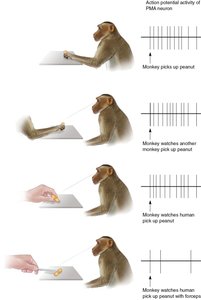
Mirror Neurons
Function and Relevance
Mirror neurons in the PMA fire both when an action is performed and when the same action is observed, suggesting a neural basis for understanding actions and intentions of others. These neurons may contribute to empathy and social cognition.
Specificity: Each mirror neuron has specific movement preferences.
Human relevance: Likely part of a system for understanding actions and intentions in humans.
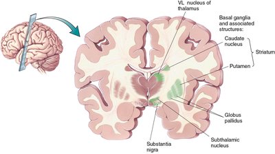
The Basal Ganglia
Structure and Function
The basal ganglia are a group of subcortical nuclei at the base of the forebrain, essential for normal brain function and behavior. They play a key role in voluntary motor control, procedural learning, routine behaviors, cognition, and emotion.
Main components: Striatum, globus pallidus, ventral pallidum, substantia nigra, subthalamic nucleus.
Motor loop: Controls selection and initiation of willful movements via a loop from cortex to basal ganglia to thalamus and back to cortex.
Basal Ganglia Disorders
Parkinson’s disease: Degeneration of dopaminergic neurons in the substantia nigra, leading to hypokinesia, bradykinesia, akinesia, rigidity, and tremors. L-dopa treatment can alleviate symptoms temporarily.
Huntington’s disease: Inherited disorder causing loss of neurons in the striatum and globus pallidus, resulting in chorea, hyperkinesia, dyskinesias, dementia, and personality disorder.
Initiation and Coding of Movement in M1
Motor Cortex and Neuronal Coding
The SMA is heavily interconnected with M1. Electrical stimulation of M1 causes contraction of specific muscle groups. Many neurons in M1 encode the force and direction of movement, and the collective activity of these neurons determines movement direction (population coding).
Population vectors: Direction of movement is determined by tallying and averaging the activity of many neurons.
The Cerebellum
Anatomy and Function
The cerebellum is divided into the vermis (midline region, axial musculature) and cerebellar hemispheres (limb movements). It is essential for the proper execution of planned, voluntary, multi-joint movements, and for sequencing muscle contractions.
Granule cells: Excitatory neurons in the cerebellar cortex.
Purkinje cells: Inhibitory neurons, largest in the cerebellar cortex.
Cerebellar lesions: Cause ataxia (uncoordinated movements), dyssynergia (loss of smooth multijoint movement), and dysmetria (overshoot/undershoot targets).
Summary Table: Major Descending Spinal Tracts
Pathway | Origin | Function |
|---|---|---|
Corticospinal tract | Motor cortex | Fine movements of arms and fingers |
Rubrospinal tract | Red nucleus | Fine movements (reduced in humans) |
Vestibulospinal tract | Vestibular nuclei | Posture and balance |
Tectospinal tract | Superior colliculus | Reflex postural movements |
Pontine reticulospinal tract | Reticular formation (pons) | Enhances antigravity reflexes |
Medullary reticulospinal tract | Reticular formation (medulla) | Inhibits extensor muscles |
Key Equations
Population Vector Calculation:
Where is the firing rate of neuron , and is its preferred direction vector.
Additional info: The baseball pitcher example is used throughout to illustrate the integration of motor planning, execution, and sensory feedback in complex voluntary movements. The notes cover the biological psychology topic, specifically the neural control of movement, relevant for college-level psychology students.
