 Back
BackChapter 2: Communication Within the Nervous System – Biological Psychology Study Notes
Study Guide - Smart Notes
 Tailored notes based on your materials, expanded with key definitions, examples, and context.
Chapter 2: Communication Within the Nervous System
Introduction
This chapter explores the structure and function of the nervous system, focusing on how neurons communicate, the role of glial cells, and the physiological basis of neural signaling. Understanding these processes is fundamental to biological psychology and the study of behavior.
The Nervous System: Structure and Function
Overview of the Nervous System
Nervous System: The body's primary communication network, consisting of the central nervous system (CNS) and peripheral nervous system (PNS).
Central Nervous System (CNS): Composed of the brain and spinal cord; processes and interprets sensory information and issues instructions.
Peripheral Nervous System (PNS): Connects the CNS to the rest of the body; includes sensory and motor neurons.
Cells of the Nervous System
Neurons: The Basic Unit
Neurons are specialized cells that transmit information throughout the nervous system via electrical and chemical signals.
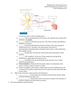
Cell Body (Soma): Contains the nucleus and organelles; integrates incoming signals.
Dendrites: Branch-like structures that receive messages from other neurons.
Axon: Long, slender projection that transmits electrical impulses away from the cell body.
Axon Terminals: Endings where neurotransmitters are released to communicate with other neurons or muscles.
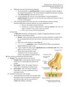
Glial Cells: Support and Insulation
Glial cells provide structural and functional support for neurons. They are essential for maintaining homeostasis, forming myelin, and protecting the brain.
Oligodendrocytes: Found in the CNS; can myelinate multiple axons.
Schwann Cells: Found in the PNS; myelinate a single axon.
Myelin Sheath: Insulating layer that speeds up neural transmission.
Neural Communication
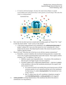
The Resting Membrane Potential
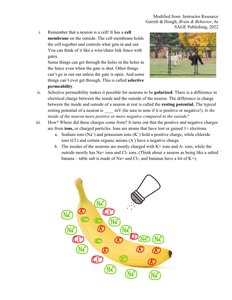
Neurons maintain a difference in electrical charge across their membrane, known as the resting membrane potential (typically around -70 mV).
Polarization: The inside of the neuron is more negative than the outside.
Sodium-Potassium Pump: Actively transports Na+ out and K+ into the cell, maintaining the resting potential.
Action Potentials: The Nerve Impulse
An action potential is a rapid, temporary change in membrane potential that travels along the axon, allowing neurons to communicate.
Threshold: The minimum depolarization needed to trigger an action potential (usually around -55 mV).
Depolarization: Na+ channels open, Na+ rushes in, making the inside more positive.
Repolarization: K+ channels open, K+ exits, restoring negativity inside.
Hyperpolarization: The membrane potential becomes more negative than resting potential before stabilizing.
Propagation: The action potential moves down the axon in a wave-like fashion.
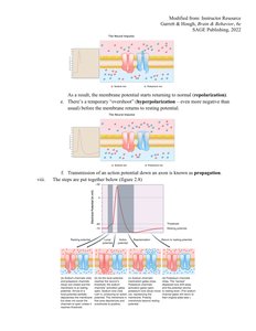
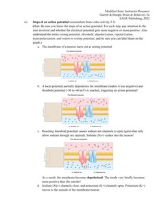
Steps of an Action Potential
The neuron is at resting potential (about -70 mV).
A stimulus causes local depolarization, reaching threshold.
Voltage-gated Na+ channels open, Na+ enters, causing rapid depolarization.
Na+ channels close, K+ channels open, K+ exits, repolarizing the cell.
Temporary hyperpolarization occurs before returning to resting potential.
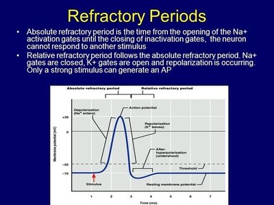
Refractory Periods
After an action potential, the neuron experiences refractory periods during which it cannot fire another action potential or requires a stronger stimulus to do so.
Absolute Refractory Period: No new action potential can be initiated.
Relative Refractory Period: A stronger-than-normal stimulus is required to initiate another action potential.
Synaptic Transmission
Chemical Synapses
Neurons communicate with each other at synapses, where neurotransmitters are released from the presynaptic neuron and bind to receptors on the postsynaptic neuron.
Excitatory Postsynaptic Potentials (EPSPs): Increase the likelihood of an action potential in the postsynaptic neuron.
Inhibitory Postsynaptic Potentials (IPSPs): Decrease the likelihood of an action potential.
Summation: The combined effect of multiple EPSPs and IPSPs determines whether the postsynaptic neuron will fire.
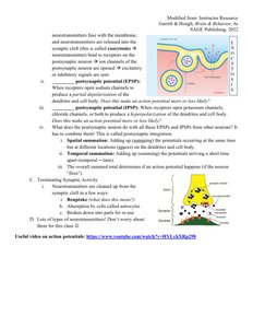
Termination of Synaptic Activity
Neurotransmitters are removed from the synaptic cleft by reuptake, enzymatic degradation, or diffusion.
Key Terms and Concepts
Neuron: Basic unit of the nervous system, specialized for communication.
Glial Cell: Support cell in the nervous system.
Action Potential: Electrical impulse that travels down the axon.
Synapse: Junction between two neurons where communication occurs.
Neurotransmitter: Chemical messenger released at synapses.
Myelin Sheath: Insulating layer around axons that increases signal speed.
Summary Table: Differences Between Oligodendrocytes and Schwann Cells
Cell Type | Location | Function | Number of Axons Myelinated |
|---|---|---|---|
Oligodendrocyte | CNS | Myelination | Multiple (up to 50) |
Schwann Cell | PNS | Myelination | One |
Key Equations
Nernst Equation (for equilibrium potential):
Resting Membrane Potential (Goldman-Hodgkin-Katz equation):
Additional info:
These notes are based on a study guide for a college-level biological psychology course, focusing on neural communication and the physiological basis of behavior.
