 Back
BackContractile Tissue: Muscle Structure and Function in Biological Psychology
Study Guide - Smart Notes
 Tailored notes based on your materials, expanded with key definitions, examples, and context.
Contractile Tissue: Muscle
Introduction to Muscle Tissue
Muscle tissue is a specialized contractile tissue essential for movement, posture, heat production, and internal organ function. It is composed of three main types: skeletal, cardiac, and smooth muscle, each with unique structural and functional properties. Muscle cells respond to chemical, electrical, and mechanical stimuli by activating a contractile mechanism involving the proteins myosin and actin.
Skeletal muscle: Voluntary, responsible for movement and posture (about 40% of body mass).
Cardiac muscle: Involuntary, found only in the heart (about 10% of body mass).
Smooth muscle: Involuntary, found in organs and blood vessels (about 10% of body mass).
Functions of Muscle Tissue
Muscle tissue serves several critical functions in the body:
Movement: Skeletal muscles pull on bones; smooth muscles move substances within organs; cardiac muscle pumps blood.
Posture & Stability: Muscles stabilize joints and maintain body position.
Support: Muscles support internal organs and shape the body.
Heat Production: Muscle activity generates heat, maintaining body temperature.
Circulation: Smooth muscle regulates blood flow; cardiac muscle pumps blood.
Protection: Muscles shield bones and organs.
Properties of Muscle Tissue
Muscle tissue exhibits five key properties:
Excitability: Ability to respond to stimuli (e.g., neurotransmitters).
Conductivity: Ability to propagate electrical signals.
Contractility: Ability to shorten and generate force.
Extensibility: Ability to stretch without damage.
Elasticity: Ability to return to original shape after stretching.
Types of Muscle Tissue
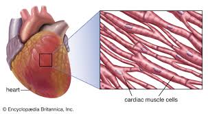
Cardiac Muscle
Cardiac muscle, or myocardium, is a specialized, involuntary, and striated muscle found exclusively in the heart. It contracts rhythmically to pump blood throughout the body and is highly resistant to fatigue due to abundant mitochondria and aerobic metabolism.
Structure: Branched, cylindrical, striated cells (cardiomyocytes) with a single central nucleus.
Intercalated Discs: Specialized junctions (gap junctions) connect cells for rapid, synchronized contraction.
Function: Responsible for rhythmic heartbeat, regulated by the sinoatrial (SA) node.
Regulation: Controlled by the autonomic nervous system, intrinsic pacemaker cells, and hormones.
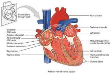
Sinoatrial (SA) Node
The SA node is the heart's natural pacemaker, located in the right atrium. It generates electrical impulses (action potentials) that initiate each heartbeat, setting the rhythm and rate. The autonomic nervous system modulates its activity, increasing heart rate during activity and decreasing it during rest.
Location: Upper wall of the right atrium.
Function: Initiates electrical cascade for heartbeat.
Clinical Significance: Dysfunction can cause bradycardia or arrhythmias.
Comparison with Other Muscle Types
Cardiac vs. Skeletal: Cardiac is involuntary and branched; skeletal is voluntary and linear.
Cardiac vs. Smooth: Cardiac is striated; smooth muscle is non-striated.
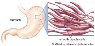
Smooth Muscle
Smooth muscle is involuntary, non-striated, and spindle-shaped. It is found in the walls of hollow organs, blood vessels, and airways, facilitating functions such as digestion, blood pressure regulation, and pupil dilation.
Structure: Fusiform (spindle-shaped), non-striated cells with a single central nucleus.
Contraction: Slow, sustained (tonic) or rhythmic (phasic) contractions.
Regulation: Controlled by the autonomic nervous system, hormones, and local chemical signals.
Types: Single-unit (visceral) and multi-unit smooth muscle.
Primary Locations and Functions
Digestive Tract: Drives peristalsis.
Cardiovascular System: Regulates blood pressure and flow.
Respiratory System: Controls airway diameter.
Urinary System: Manages urine flow.
Reproductive System: Contracts during pregnancy.
Clinical Significance
Disruption can cause asthma, hypertension, and gastrointestinal motility disorders.
Skeletal Muscle
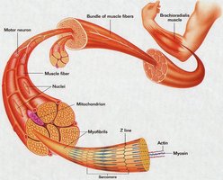
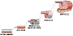
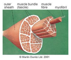
Skeletal muscle is voluntary, striated, and composed of long, cylindrical, multinucleated fibers. It is responsible for movement, posture, and heat production. Each muscle fiber is surrounded by a cell membrane (sarcolemma) and contains myofibrils made up of myofilaments (actin and myosin).
Structure: Long, cylindrical, multinucleated fibers; nuclei at periphery.
Organization: Muscle fibers grouped into bundles (fascicles), which form the muscle.
Contraction: Requires nervous stimulation; lacks syncytial bridges between cells.
Muscle Proteins and Contractile Mechanism
Myofibrils and Myofilaments
Myofibrils are composed of repeating units called sarcomeres, which contain thick (myosin) and thin (actin) filaments. These filaments are organized to facilitate contraction.
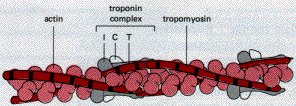
Actin: Thin filament; primary protein in muscle contraction.
Myosin: Thick filament; motor protein with ATPase activity.
Tropomyosin: Covers actin's active sites.
Troponin: Regulatory protein with three subunits (I, T, C).
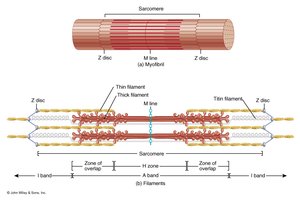
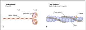
Sliding Filament Theory
The sliding filament theory explains muscle contraction: thin filaments slide past thick filaments, shortening the sarcomere. This process is driven by cross-bridge cycling between actin and myosin, regulated by calcium ions and ATP.
Myosin heads: Bind actin and hydrolyze ATP.
Actin: Contains myosin-binding sites, covered by tropomyosin at rest.
Troponin C: Binds calcium, causing tropomyosin to move and expose binding sites.
Excitation-Contraction Coupling Steps
Action potential propagates to T tubules.
Voltage-sensitive Ca2+ gates open on sarcoplasmic reticulum.
Ca2+ released into muscle fiber cytoplasm.
Ca2+ binds troponin C, causing conformational change.
Tropomyosin moves, exposing myosin-binding sites on actin.
Cross-bridge cycling: myosin binds actin, hydrolyzes ATP, generates force.
Relaxation: Ca2+ reaccumulated in sarcoplasmic reticulum, tropomyosin blocks binding sites, muscle relaxes.

Muscle Fiber Physiology and Neuromuscular Junction
Muscle Fiber Excitability
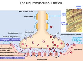
Muscle fibers can be excited chemically, electrically, and mechanically. Each fiber receives innervation from a motor neuron at the neuromuscular junction (NMJ), where neurotransmitter acetylcholine (ACh) initiates contraction.
Neuromuscular Junction (NMJ)
The NMJ is the synapse between a motor neuron and a muscle fiber. It is essential for translating neural signals into muscle contraction.
Synaptic end bulbs: Contain vesicles of ACh.
Motor end plate: Contains ACh receptors.
Steps: AP arrives, Ca2+ influx, ACh release, ACh binds receptors, Na+ influx, muscle AP, contraction, ACh breakdown.
Pharmacology of the NMJ
Botulinum toxin: Blocks ACh release, causing paralysis.
Curare: Blocks ACh receptors, causing paralysis.
Neostigmine: Inhibits ACh breakdown, strengthens contractions in myasthenia gravis.
Muscle Pathophysiology
Rigor Mortis
Rigor mortis is the postmortem rigidity of muscles due to ATP depletion, preventing Ca2+ reuptake and myosin detachment from actin, resulting in irreversible contraction.
Tetanus (Tetanic Contraction)
Tetanic contraction occurs when repeated stimulation prevents muscle relaxation, leading to sustained, maximal tension. Complete tetanus has no relaxation between stimuli; incomplete tetanus has partial relaxation.
Muscle Type | Structure | Control | Location | Function |
|---|---|---|---|---|
Skeletal | Striated, multinucleated, cylindrical | Voluntary | Attached to bones | Movement, posture |
Cardiac | Striated, branched, single nucleus | Involuntary | Heart | Pumping blood |
Smooth | Non-striated, spindle-shaped, single nucleus | Involuntary | Organs, vessels | Internal movement |
Example: Skeletal muscle contracts to move limbs; cardiac muscle contracts to pump blood; smooth muscle contracts to move food through the digestive tract.
Additional info: These notes expand on the original content with academic context, definitions, and examples to provide a comprehensive overview suitable for psychology students studying biological psychology and muscle physiology.
