 Back
BackSensation and Perception: The Visual, Auditory, Vestibular, Somatosensory, Olfactory, and Gustatory Systems
Study Guide - Smart Notes
 Tailored notes based on your materials, expanded with key definitions, examples, and context.
The Visual System
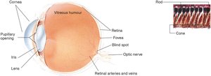
Major Parts of the Human Eye
The human eye is a complex organ responsible for detecting light and converting it into neural signals for the brain to interpret as vision. Understanding the structure and function of each part is essential for grasping how visual information is processed.
Cornea: The transparent, protective outer membrane that bends light rays inward, helping to focus them.
Sclera: The white, protective outer layer of the eye.
Iris: The colored part of the eye, controlling the size of the pupil and thus the amount of light entering the eye.
Pupil: The opening in the center of the iris through which light enters the eye.
Lens: A transparent structure that changes shape (accommodation) to focus light onto the retina.
Retina: The light-sensitive membrane at the back of the eye where photoreceptors (rods and cones) are located.
Fovea: The central region of the retina responsible for sharp central vision (visual acuity).
Optic Nerve: Transmits visual information from the retina to the brain.
Vitreous Humour: The clear gel that fills the space between the lens and the retina.
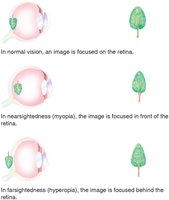
Focusing and Visual Acuity
The lens focuses light onto the retina, and its curvature changes to adjust for objects at different distances. Glasses or contact lenses can correct focusing problems such as myopia (nearsightedness) and hyperopia (farsightedness).
Myopia: Image is focused in front of the retina; distant objects appear blurry.
Hyperopia: Image is focused behind the retina; close objects appear blurry.
Normal Vision: Image is focused directly on the retina.
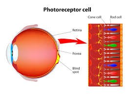
Photoreceptors: Rods and Cones
The retina contains two main types of photoreceptors: rods and cones. These cells convert light into neural signals.
Rods: Sensitive to low light; responsible for black-and-white vision and peripheral vision. More numerous in the periphery of the retina.
Cones: Responsible for color vision and fine detail; concentrated in the fovea. Do not function well in dim light.
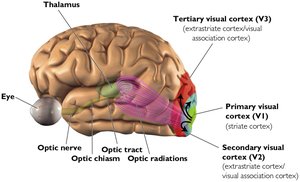
Visual Pathways to the Brain
Visual information is transmitted from the retina through the optic nerve to the visual cortex at the back of the brain. The brain processes and interprets these signals, flipping the upside-down image projected on the retina to the correct orientation.
Primary Visual Cortex (V1): The first cortical area to receive visual input.
Secondary and Tertiary Visual Cortices (V2, V3): Further process and interpret visual information.
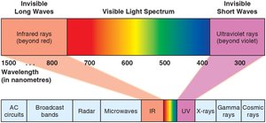
The Visible Spectrum
Humans can detect only a narrow range of the electromagnetic spectrum, known as the visible spectrum. Wavelength determines color, and amplitude determines brightness.
Wavelength: Determines the perceived color of light.
Amplitude: Determines the perceived brightness of light.
The Auditory System
Physical Properties of Sound
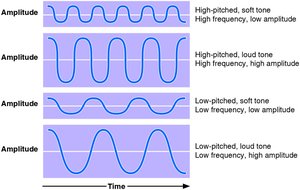
Sound is a form of energy that travels in waves. The auditory system detects and interprets these waves as sound.
Frequency (Hz): Number of cycles per second; determines pitch.
Amplitude (dB): Height of the sound wave; determines loudness.
Timbre: Quality of sound that distinguishes different sources.
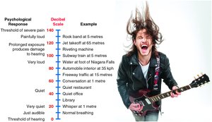
Decibel Scale and Hearing Safety
Loudness is measured in decibels (dB). Each increase of 10 dB represents a tenfold increase in loudness. Prolonged exposure to sounds above 130 dB can cause immediate hearing damage.
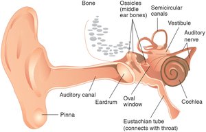
Anatomy of the Ear
The ear is divided into three main sections: outer, middle, and inner ear. Each part plays a role in transmitting and amplifying sound waves.
Outer Ear: Includes the pinna and auditory canal; collects and channels sound waves.
Middle Ear: Contains the ossicles (hammer, anvil, stirrup) that amplify vibrations.
Inner Ear: Contains the cochlea, where sound waves are converted into neural signals by hair cells.
Theories of Hearing
Two main theories explain how we perceive pitch:
Place Theory: Different frequencies stimulate different places along the basilar membrane in the cochlea. Explains perception of high-frequency sounds.
Frequency Theory: The rate of nerve impulses matches the frequency of a tone, explaining perception of low-frequency sounds.
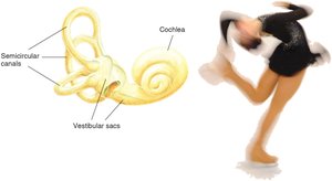
The Vestibular System
Balance and Spatial Orientation
The vestibular system is responsible for our sense of balance and spatial orientation. It detects head movement and position, sending information to the brain to help maintain balance.
Vestibular Sacs: Detect head position, especially when not upright.
Semicircular Canals: Detect rotational movements of the head.
Connections: Information is sent to the brainstem, cerebellum, and amygdala.
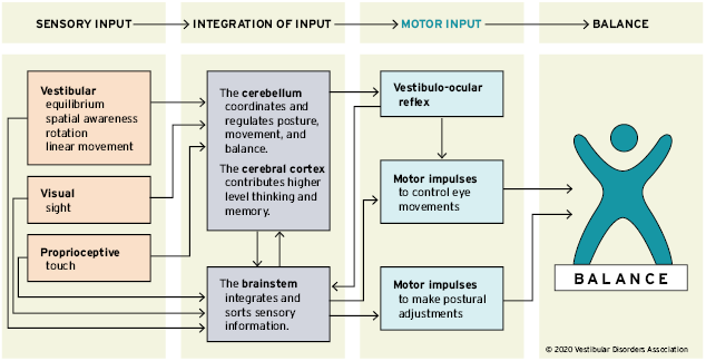
Sensing Balance and Movement
Movement of fluid in the semicircular canals bends hair-cell receptors, which send signals to the brain about head rotation and movement.
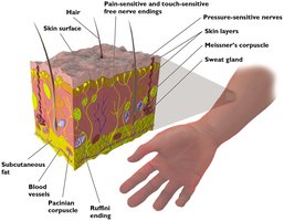
The Somatosensory System
Sense of Touch
The somatosensory system detects stimuli such as pressure, temperature, and injury through specialized nerve endings in the skin.
Mechanoreceptors: Specialized nerve endings that respond to mechanical pressure or distortion.
Free Nerve Endings: Detect pain and temperature.
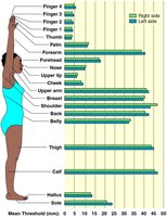
Measuring Touch Acuity: Two-Point Threshold
The two-point threshold measures the minimum distance at which two points of contact can be felt as separate. Sensitivity varies across different body parts.
Fingers and thumb: Highest sensitivity (lowest threshold).
Calves: Lowest sensitivity (highest threshold).
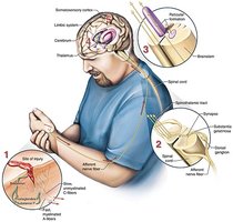
Pain Sensation and the Gate Control Model
Pain is detected by nerve endings near the skin's surface. The Gate Control Model suggests that neural mechanisms in the spinal cord regulate the perception of pain, which can be influenced by cognitive and emotional factors.
Small fibers: Transmit pain signals.
Large fibers: Inhibit pain signals.
Phantom pain: Pain perceived in a missing limb.
Acute vs. Chronic Pain
Pain can be classified as acute or chronic based on its duration and underlying cause.
Acute Pain: Short-term, identifiable source, improves with rest.
Chronic Pain: Lasts longer than 3 months, may become a disease itself, requires careful management.
The Role of the Brain in Pain
The brain interprets pain signals and can modulate them through inhibitory systems or the release of endorphins, which are natural painkillers.
Endorphins: Chemicals produced by the body that reduce pain and promote well-being (e.g., runner's high).
Olfactory System: Sense of Smell
Olfaction
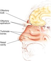
Olfaction is the sense of smell, a chemical sense that detects airborne molecules.
Olfactory Epithelium: Tissue at the top of the nasal cavity containing millions of smell receptor cells.
Olfactory Bulbs: Structures above the nasal cavity where smell sensations are first processed in the brain.
Olfactory Nerve: Transmits signals from the olfactory receptors to the brain.
Gustatory System: Sense of Taste
Gustation
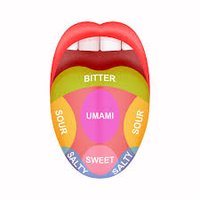
The gustatory system is responsible for the sense of taste, detecting chemicals dissolved in saliva.
Basic Tastes: Sweet, sour, salty, bitter, and umami (savory).
Taste Buds: Located on papillae on the tongue; each contains 60-100 receptor cells and is replaced every 10 days.
Variation in Taste Sensitivity: Non-tasters, medium tasters, and supertasters differ in the number of taste buds.
Interaction of Smell and Taste
Olfaction and gustation work together to create the perception of flavor. Both are considered chemical senses and their signals converge in the orbitofrontal cortex of the brain.
