 Back
BackSensation and Perception: Visual Stimuli and Color Vision
Study Guide - Smart Notes
 Tailored notes based on your materials, expanded with key definitions, examples, and context.
Visual Stimuli
Nature of Visual Stimuli
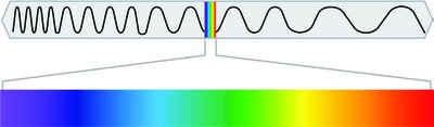
The stimulus for vision is light, specifically electromagnetic energy within the visible spectrum (approximately 380 to 700 nanometers). The physical properties of light waves determine the qualities we perceive visually.
Wavelength: The distance between successive peaks of a wave; determines hue (color).
Amplitude: The height of the wave; determines brightness.
Complexity: The number of different wavelengths present; determines saturation (purity of color).
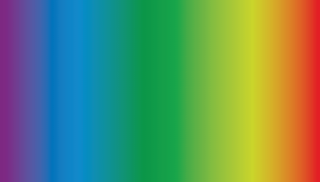
Visible light spectrum is a small portion of the electromagnetic spectrum, ranging from violet (shortest wavelength) to red (longest wavelength).
Visual Anatomy
Structure of the Eye
The human eye is a complex organ that focuses light and converts it into neural signals for the brain to interpret.
Cornea: The transparent, protective outer layer that bends (refracts) light as it enters the eye.
Pupil: The opening in the center of the iris that allows light to enter the eye.
Iris: The colored part of the eye surrounding the pupil; controls the size of the pupil.
Lens: A transparent structure that changes shape (accommodation) to focus light on the retina.
Retina: The neural tissue lining the back of the eyeball; contains photosensitive cells (rods and cones) that detect light.
Accommodation is the process by which the lens changes shape to focus on objects at different distances. This ability decreases with age, often requiring reading glasses.

Pathway of Light Through the Eye
Light passes through the following structures in order:
Cornea
Pupil (regulated by the iris)
Lens
Retina (where neural signals are generated)
Photosensitive Cells
The retina contains two main types of photosensitive cells:
Rods: Sensitive to low light; responsible for night vision and peripheral vision. Located mainly in the periphery of the retina. Enable achromatic (black-and-white) vision. There are many rods in the retina.
Cones: Sensitive to bright light and color; concentrated in the fovea (center of the retina). Responsible for high visual acuity and color vision. There are fewer cones than rods, and three types of cones exist, each sensitive to different wavelengths (colors).
The fovea is the center of the retina and contains a high density of cones, allowing for sharp central vision. The blind spot is the area where the optic nerve exits the eye; it contains no rods or cones.

Color Vision
Trichromatic Theory
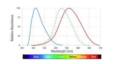
The trichromatic theory explains the first stage of color vision, which occurs in the retina. According to this theory, there are three types of cones, each sensitive to a different range of wavelengths (short, medium, and long, corresponding roughly to blue, green, and red).
Each type of cone responds maximally to a specific wavelength.
Color perception arises from the relative activation of these three types of cones.
This theory explains how we can perceive a wide range of colors by mixing signals from the three cone types.
Opponent Process Theory
The opponent process theory describes the second stage of color processing, which occurs in the nervous system beyond the retina. This theory proposes that color is processed in terms of opposing pairs:
Red vs. Green
Blue vs. Yellow
Black vs. White
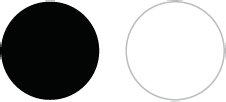
Opponent process cells are excited by one color in the pair and inhibited by the other. This explains phenomena such as afterimages, where staring at one color for a long time results in seeing its opposite when looking away.
Colorblindness
Colorblindness is a condition where one or more types of cones are nonfunctional. The most common type is deuteranopia, where the green-sensitive cones are absent or not working. This affects the ability to distinguish between certain colors, especially reds and greens.
Summary Table: Properties of Light and Perception
Wave Property | Perceived Property |
|---|---|
Wavelength | Hue (Color) |
Amplitude | Brightness |
Complexity | Saturation |
Key Equations
Relationship between wavelength and frequency: where is the speed of light, is wavelength, and is frequency.
Applications and Examples
Looking at a colorful painting relies on cones (color vision and high acuity).
Walking in dim light relies on rods (sensitivity to low light).
Peripheral vision is mainly mediated by rods.
Colorblindness can be diagnosed by testing responses to different colors.
