 Back
BackSensation and Perception: Visual Stimuli and Color Vision
Study Guide - Smart Notes
 Tailored notes based on your materials, expanded with key definitions, examples, and context.
Visual Stimuli
Introduction to Visual Stimuli
The process of vision begins with the detection of visual stimuli, which are forms of electromagnetic energy. The human visual system is sensitive to a narrow range of wavelengths known as the visible light spectrum, spanning approximately 380 to 700 nanometers (nm).
Stimulus for vision: Light waves (electromagnetic radiation within the visible spectrum).
Wave properties: The physical characteristics of light waves determine the qualities we perceive.
Wavelength: Determines hue (color).
Amplitude: Determines brightness.
Complexity: Determines saturation (purity of color).
Visible light spectrum: The range of light wavelengths visible to the human eye, from violet (shortest wavelength) to red (longest wavelength).
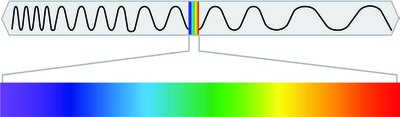
Wave Properties and Perceived Qualities
Wavelength: How long the wave is; perceived as hue (color).
Amplitude: How tall the wave is; perceived as brightness.
Complexity: How many different wavelengths are present; perceived as saturation.
Example: A pure red light (single wavelength) is highly saturated, while white light (many wavelengths) is less saturated.
Visual Anatomy
Structure of the Eye
The eye is a complex organ that focuses light and converts it into neural signals for the brain to interpret.
Cornea: The transparent, protective outer layer that bends (refracts) light as it enters the eye.
Pupil: The opening in the center of the iris that allows light to enter the eye.
Iris: The colored part of the eye, which controls the size of the pupil.
Lens: A flexible structure that changes shape (accommodation) to focus light onto the retina.
Retina: The light-sensitive layer at the back of the eye containing photoreceptor cells (rods and cones).
Accommodation: The process by which the lens changes shape to focus on objects at different distances. This ability decreases with age, often requiring reading glasses.

Pathway of Light Through the Eye
Light enters the cornea, passes through the pupil (regulated by the iris), is focused by the lens, and finally reaches the retina where it is converted into neural signals.
Order of structures: Cornea → Pupil → Lens → Retina → Neural signal
Photosensitive Cells
The retina contains two main types of photoreceptor cells: rods and cones.
Rods: Rod-shaped cells that respond to low light levels (scotopic vision), are located mainly in the periphery of the retina, and are responsible for detecting movement and shapes. There are many rods, but they do not detect color.
Cones: Cone-shaped cells that respond to bright light (photopic vision), are concentrated in the fovea (center of the retina), and are responsible for color vision and high visual acuity. There are fewer cones, and they are divided into three types, each sensitive to different wavelengths (red, green, blue).
Fovea: The central region of the retina with a high density of cones, providing sharp central vision.
Blind spot: The point where the optic nerve exits the eye; contains no photoreceptors, resulting in a gap in the visual field.

Color Vision
Trichromatic Theory
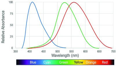
The trichromatic theory explains color vision at the level of the retina. It proposes that there are three types of cones, each sensitive to a specific range of wavelengths (short, medium, long), corresponding to blue, green, and red light.
Three types of cones: Each type is maximally sensitive to a different part of the visible spectrum.
Color perception: The brain interprets the relative activation of these cones to produce the experience of color.
Limitation: This theory cannot explain all aspects of color vision, such as afterimages and certain types of color blindness.
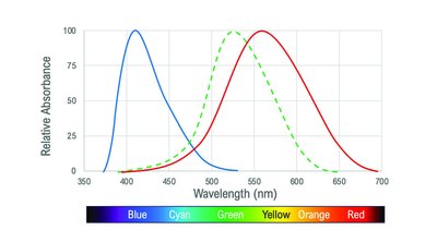
Opponent Process Theory
The opponent process theory describes a later stage of color processing, occurring in the nervous system beyond the retina. It suggests that color perception is controlled by the activity of two opponent systems: red-green, blue-yellow, and black-white.
Opponent pairs: Red/green, blue/yellow, black/white.
Mechanism: Opponent process cells are excited by one color and inhibited by its opponent color.
Afterimages: Staring at one color for a prolonged period can lead to the perception of its opponent color when looking away (e.g., seeing green after staring at red).
Colorblindness
Colorblindness is a condition in which one or more types of cones are nonfunctional. The most common type is deuteranopia, where the green-sensitive cones are absent or nonfunctional.
Deuteranopia: Individuals have difficulty distinguishing between red and green hues.
Diagnosis: Based on which colors are seen well and which are not, the affected cone type can be identified.
Summary Table: Properties of Light and Perception
Wave Property | Perceived Quality |
|---|---|
Wavelength | Hue (Color) |
Amplitude | Brightness |
Complexity | Saturation |
Summary Table: Photoreceptor Comparison
Photoreceptor | Location | Function | Color Sensitivity | Number |
|---|---|---|---|---|
Rods | Periphery of retina | Low light, movement | No | Many |
Cones | Fovea (center of retina) | Bright light, color, detail | Yes (3 types) | Few |
Additional info: The visual system integrates information from both trichromatic and opponent process mechanisms to produce the rich experience of color vision. Disorders such as colorblindness provide insight into the functioning and limitations of these systems.
