 Back
BackThe Biological Basis of Behavior: Brain Structure and Function
Study Guide - Smart Notes
 Tailored notes based on your materials, expanded with key definitions, examples, and context.
The Brain: Introduction and Importance
Why Study the Brain?
The brain is the central organ of the nervous system and is responsible for cognition, emotion, and consciousness. Understanding the brain helps establish the reality of psychological phenomena and deepens our knowledge of mental processes.
Cognition: The mental processes involved in acquiring knowledge and understanding.
Emotion: The experience and expression of feelings.
Consciousness: Awareness of self and environment.
Major Brain Structures and Their Functions
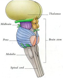
Brainstem
The brainstem is the oldest part of the brain, connecting to the spinal cord and responsible for automatic survival functions.
Medulla: Controls heart rate, breathing, swallowing, digestion, salivating, coughing, and sneezing.
Pons: Acts as a relay station between the cerebellum and cerebrum; involved in respiration and the sleep-wake cycle.
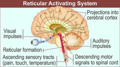
Reticular Formation: Controls arousal and consciousness; damage can cause coma.
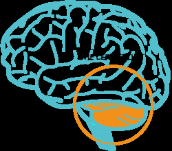
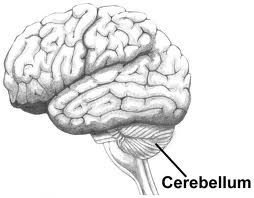
Cerebellum
The cerebellum sits behind the top portion of the brainstem and contains about 90% of the brain's neurons. It is involved in fine motor control, coordination, balance, and procedural memory.
Damage can cause problems with balance, coordination, and judging distances.
Alcohol significantly impairs cerebellar function.
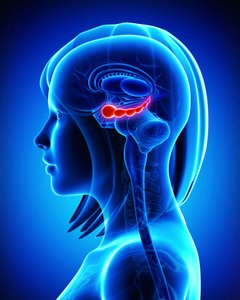
Thalamus
The thalamus sits atop the brainstem and acts as the brain's sensory control center, processing all senses except smell. It relays incoming sensory information to the cortex for further processing.
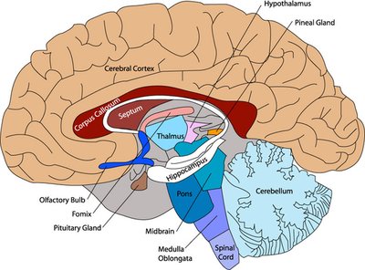
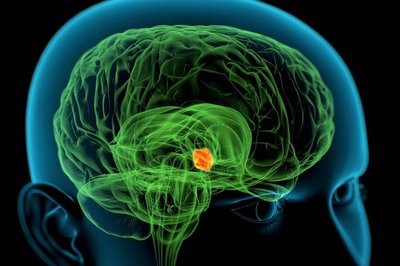
Limbic System
The limbic system is a group of brain structures involved in processing emotion, motivation, drives (such as food and aggression), and memory.
Amygdala: Processes emotions, especially fear and aggression; triggers fight-or-flight responses.
Hippocampus: Involved in the processing and storage of new memories, spatial memory, and the transfer of information from short-term to long-term memory.
Hypothalamus: Regulates the autonomic nervous system, body temperature, hunger, thirst, and the four F's: feeding, fighting, fleeing, and mating.
Corpus Callosum
The corpus callosum is a large band of neural fibers connecting the left and right brain hemispheres, allowing communication between them. Severing the corpus callosum results in a "split brain," often used as a treatment for severe epilepsy.
Cerebral Cortex
The cerebral cortex is the outermost layer of the brain, responsible for higher-order functions such as perception, thought, and decision-making. It is divided into two hemispheres, each with four lobes: frontal, parietal, temporal, and occipital.
Contains approximately 30 billion nerve cells and 270 billion glial cells.
Wrinkles (gyri and sulci) increase surface area for more neural connections.
Lobes of the Cerebral Cortex and Their Functions
Frontal Lobes
The frontal lobes are involved in executive functions such as judgment, planning, reasoning, and problem-solving. The motor cortex, located here, controls voluntary movement.
Prefrontal Cortex: Responsible for moral judgment and personality.
Broca’s Area: Involved in speech production; damage leads to Broca’s aphasia (difficulty producing speech).
Case Study: Phineas Gage
Phineas Gage suffered damage to his prefrontal cortex, resulting in profound personality changes and loss of moral judgment.
Parietal Lobes
The parietal lobes process sensory signals from the body, including touch, pressure, temperature, and pain. They are also involved in spatial awareness and body movement.
Somatosensory Cortex: Registers and processes body touch and movement sensations.
Temporal Lobes
The temporal lobes are involved in hearing, language processing, and memory. They contain the main auditory cortex and are essential for understanding language.
Wernicke’s Area: Responsible for language comprehension; damage leads to Wernicke’s aphasia (difficulty understanding and producing meaningful language).
Right Fusiform Gyrus: Allows recognition of human faces; damage can cause prosopagnosia (face blindness).
Occipital Lobes
The occipital lobes process visual information from the eyes. Damage can result in blindness or visual disturbances.
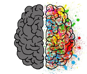
Brain Lateralization and Plasticity
Brain Lateralization
Brain lateralization refers to the specialization of certain functions to either the left or right hemisphere. The brain is cross-wired, meaning each hemisphere controls the opposite side of the body.
Plasticity and Neurogenesis
Plasticity is the brain’s ability to reorganize itself by forming new neural connections, especially after injury. Neurogenesis is the formation of new neurons, which occurs in limited brain regions (memory and smell).
Methods for Studying the Brain
Case Studies
Case studies involve in-depth analysis of individuals or groups to understand brain function. Notable examples include:
Phineas Gage: Frontal lobe damage and personality change.
Patient H.M.: Removal of hippocampus led to inability to form new memories.
Clive Wearing: Severe amnesia affecting both past memories and the ability to form new ones.
Lesion Studies
Lesion studies involve removing or damaging parts of the brain to observe resulting deficits. They help identify the functions of specific brain regions but can be limited by unintended damage to adjacent areas.
Brain Imaging Techniques
EEG (Electroencephalogram): Measures electrical activity in the brain using scalp electrodes; useful for detecting seizures and abnormalities.
PET Scan (Positron Emission Tomography): Measures metabolic activity by tracking glucose absorption; shows active brain regions.
CT Scan (Computed Tomography): Uses x-rays to examine brain structure; useful for detecting tumors and structural abnormalities.
MRI (Magnetic Resonance Imaging): Uses magnetic fields and radiofrequency pulses to produce detailed images of brain tissue; superior for soft tissue visualization.
fMRI (Functional MRI): Measures brain function by tracking blood flow and oxygen use; more precise than PET scans.
Comparison of CT and MRI
Feature | CT Scan | MRI |
|---|---|---|
Imaging Method | X-rays (radiation) | Magnetic fields & radiofrequency |
Best For | Bone, structure, tumors | Soft tissue, brain, organs |
Detail Level | Moderate | High |
Summary Table: Major Brain Structures and Functions
Structure | Main Function |
|---|---|
Medulla | Vital functions (heart rate, breathing) |
Pons | Relay station, sleep-wake cycle |
Cerebellum | Coordination, balance, procedural memory |
Thalamus | Sensory relay (except smell) |
Limbic System | Emotion, motivation, memory |
Corpus Callosum | Connects hemispheres |
Frontal Lobe | Executive functions, movement, speech |
Parietal Lobe | Sensory processing, spatial awareness |
Temporal Lobe | Hearing, language, memory |
Occipital Lobe | Vision |
Additional info: This guide expands on the provided notes with definitions, examples, and tables for clarity and exam preparation.
