 Back
BackThe Biological Perspective: Structure and Function of the Nervous System
Study Guide - Smart Notes
 Tailored notes based on your materials, expanded with key definitions, examples, and context.
The Nervous System
Overview
The nervous system is an extensive network of specialized cells that carry information to and from all parts of the body. Neuroscience is the scientific study of the structure and function of neurons, nerves, and nervous tissue, and their relationship to behavior and learning.
Nervous System: Coordinates actions and sensory information by transmitting signals to and from different parts of the body.
Neuroscience: Explores how neural structures and functions relate to behavior and learning.
Structure of the Neuron
Parts of a Neuron and Their Function
Neurons are the basic cells of the nervous system, responsible for receiving and sending messages. Each neuron consists of several key parts:
Dendrites: Branch-like structures that receive messages from other neurons.
Soma: The cell body, responsible for maintaining the life of the cell.
Axon: Long, tube-like structure that carries the neural message to other cells.
Axon Terminals: Rounded areas at the end of the axon branches, responsible for communicating with other nerve cells.
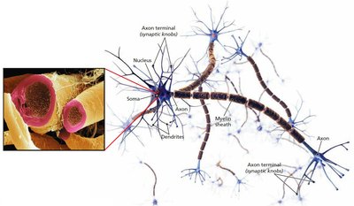
Other Types of Brain Cells
Glial Cells: Grey fatty cells that provide support for neurons, deliver nutrients, produce myelin, and clean up waste products and dead neurons.
Myelin: Fatty substance produced by glial cells that coats axons, insulating and speeding up neural impulses.
Neural Impulse: Action Potential
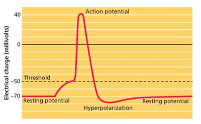
Generating the Message
Neurons communicate via electrical impulses.
Ions: Charged particles; inside the neuron are negatively charged, outside are positively charged.
Resting Potential: The state of the neuron when not firing a neural impulse.
Action Potential: The release of the neural impulse, consisting of a reversal of the electrical charge within the axon, allowing positive sodium ions to enter the cell.
All-or-None Principle: A neuron either fires completely or not at all.
Communication Between Neurons
Neurotransmitters and Synaptic Transmission
Neurons communicate with each other using chemical messengers called neurotransmitters.
Synaptic Vesicles: Sack-like structures containing neurotransmitters, found inside the axon terminal.
Neurotransmitter: Chemical released from synaptic vesicles that affects the next cell.
Synapse/Synaptic Gap: Microscopic fluid-filled space between the axon terminals of one cell and the dendrites or surface of the next cell.
Receptor Sites: Holes in the surface of dendrites or certain cells, shaped to fit only certain neurotransmitters.
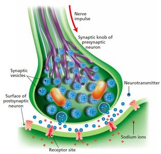
Excitatory Neurotransmitter: Causes the receiving cell to fire.
Inhibitory Neurotransmitter: Causes the receiving cell to stop firing.
Agonists: Mimic or enhance the effects of a neurotransmitter.
Antagonists: Block or reduce a cell’s response to neurotransmitters.
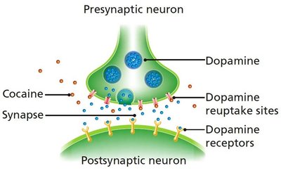
Cleaning Up the Synapse
Reuptake: Process by which neurotransmitters are taken back into the synaptic vesicles.
Enzyme: Complex protein that breaks up neurotransmitters, such as acetylcholine, for rapid muscle activity.
Major Neurotransmitters and Their Functions
Neurotransmitter | Function |
|---|---|
Acetylcholine (ACh) | Excitatory or inhibitory; involved in arousal, attention, memory, and controls muscle contractions |
Norepinephrine (NE) | Mainly excitatory; involved in arousal and mood |
Dopamine (DA) | Excitatory or inhibitory; involved in control of movement and sensations of pleasure |
Serotonin (5-HT) | Excitatory or inhibitory; involved in sleep, mood, anxiety, and appetite |
GABA | Major inhibitory neurotransmitter; involved in sleep and inhibits movement |
Glutamate | Major excitatory neurotransmitter; involved in learning, memory formation, nervous system development, and synaptic plasticity |
Endorphins | Inhibitory neural regulators; involved in pain relief |
Organization of the Nervous System
Central and Peripheral Nervous Systems
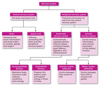
The nervous system is divided into the central nervous system (CNS) and the peripheral nervous system (PNS).
Central Nervous System (CNS): Consists of the brain and spinal cord.
Spinal Cord: Carries messages to and from the body to the brain; responsible for fast, lifesaving reflexes.
Peripheral Nervous System (PNS): All nerves and neurons outside the CNS; divided into somatic and autonomic nervous systems.
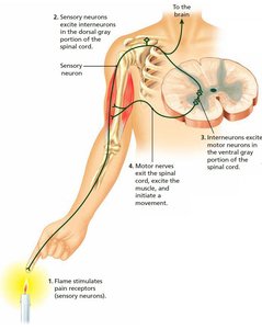
Reflex Arc and Neuroplasticity
Three Types of Neurons
Sensory Neuron (Afferent): Carries information from the senses to the CNS.
Motor Neuron (Efferent): Carries messages from the CNS to the muscles.
Interneuron: Found in the spinal cord and brain; receives information from sensory neurons and sends commands to muscles via motor neurons.
Neuroplasticity: The ability to change the structure and function of cells in response to experience or trauma.
Somatic and Autonomic Nervous Systems
Somatic Nervous System
Somatic Nervous System: Controls voluntary muscles and transmits sensory information to the CNS.
Sensory Pathway: Nerves coming from sensory organs to the CNS.
Motor Pathway: Nerves coming from the CNS to voluntary muscles.
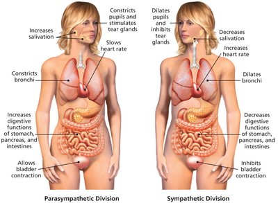
Autonomic Nervous System
Autonomic Nervous System (ANS): Controls involuntary muscles, organs, and glands.
Sympathetic Division: Reacts to stressful events and arousal (fight or flight).
Parasympathetic Division: Restores the body to normal functioning after arousal; responsible for day-to-day functioning.
The Endocrine System
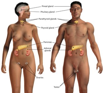
Endocrine Glands and Hormones
The endocrine system consists of glands that secrete hormones directly into the bloodstream, affecting behavior and bodily functions.
Pituitary Gland: Secretes growth hormone; influences other glands (master gland).
Pineal Gland: Secretes melatonin; regulates sleep.
Thyroid Gland: Regulates metabolism.
Pancreas: Controls blood sugar levels.
Gonads: Ovaries (female) and testes (male); regulate sexual development and reproduction.
Adrenal Glands: Secrete hormones for stress response; regulate salt intake; secondary source of sex hormones.
Stress and the Nervous System
General Adaptation Syndrome (GAS)
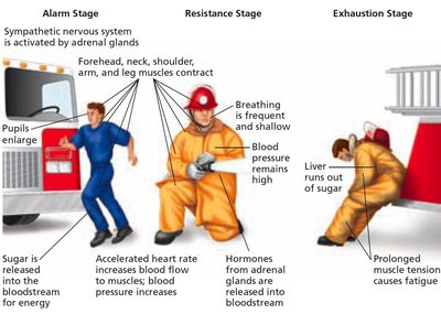
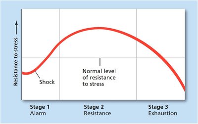
Stress impacts the autonomic nervous system and the body through three stages:
Alarm: Sympathetic nervous system is activated.
Resistance: Body remains alert and active.
Exhaustion: Body's resources are depleted, leading to fatigue.
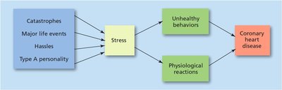
Stress and the Immune System
Immune System: Responds to disease, infection, and injury; negatively affected by stress.
Psychoneuroimmunology: Study of psychological factors on the immune system.
Heart Disease: Stress increases risk for coronary heart disease.
Diabetes: Type 2 diabetes is associated with excessive weight gain and inefficient insulin levels.
Cancer: Stress increases malfunction of natural killer (NK) cells, which suppress viruses and destroy tumor cells.
Studying the Brain
Lesioning and Brain Stimulation
Transcranial Magnetic Stimulation (TMS): Magnetic pulses applied to the cortex.
Transcranial Direct Current Stimulation (tDCS): Direct electrical stimulation of the brain.
Human Brain Damage: Clinical studies of brain injury.
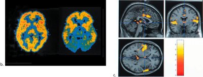
Neuroimaging Techniques
Computed Tomography (CT): Computer-controlled X-rays of the brain.
Magnetic Resonance Imaging (MRI): Uses radio waves and magnetic fields for detailed images.
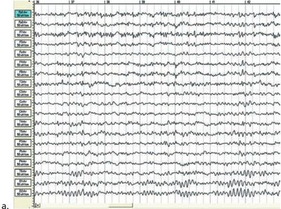
Electroencephalogram (EEG): Records electrical activity of the brain.
Positron Emission Tomography (PET): Radioactive sugar is injected; computer compiles color-coded images of brain activity.
Single Photon Emission Computed Tomography (SPECT): Similar to PET, uses different tracers.
Functional MRI (fMRI): Creates a "movie" of changes in brain activity over time.
Major Structures of the Human Brain
The Hindbrain
Medulla: Controls life-sustaining functions (breathing, heart rate).
Pons: Connects top and bottom of brain; involved in sleep, dreaming, coordination, and arousal.
Reticular Formation: Responsible for selective attention.
Cerebellum: Coordinates involuntary, rapid, fine motor movement.
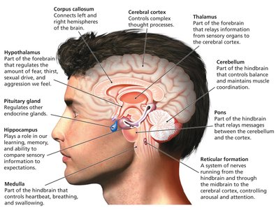
Structures Under the Cortex: The Limbic System
Limbic System: Group of structures involved in learning, emotion, memory, and motivation.
Thalamus: Relays sensory information to the cortex.
Hypothalamus: Regulates motivational behaviors (sleep, hunger, thirst, sex).
Hippocampus: Formation of long-term memories and spatial memory.
Amygdala: Responsible for fear responses and memory of fear.
Cingulate Cortex: Important for cognitive and emotional processing.
The Cortex and Cerebral Hemispheres
Parts of the Cortex Controlling Senses and Movement
Cortex: Outermost covering of the brain; responsible for higher thought processes and interpretation of sensory input.
Corticalization: Wrinkling of the cortex, increasing surface area.
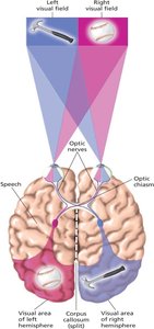
Cerebral Hemispheres: Left and right sections of the cortex, connected by the corpus callosum.
Four Lobes of the Brain
Occipital Lobe: Visual processing.
Parietal Lobe: Touch, taste, and temperature sensations.
Temporal Lobe: Hearing and meaningful speech.
Frontal Lobe: Higher mental processes, decision making, fluent speech.
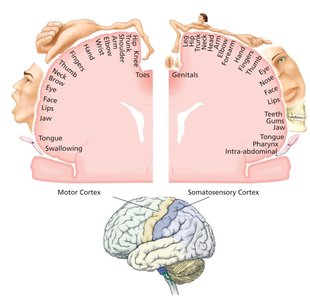
Motor Cortex: Sends motor commands to muscles.
Somatosensory Cortex: Processes information from skin and internal body receptors.
Association Areas and Higher Thought
Association Areas of Cortex
Association Areas: Coordinate and interpret information, responsible for higher mental processing.
Broca’s Aphasia: Damage to Broca’s area (left frontal lobe) causes difficulty speaking fluently.
Wernicke’s Aphasia: Damage to Wernicke’s area (left temporal lobe) causes difficulty understanding or producing meaningful language.
Spatial Neglect: Damage to right hemisphere association areas results in inability to recognize objects/body parts in the left visual field.
Split-Brain Research and Hemispheric Specialization
Differences Between Left and Right Sides of the Brain
Cerebrum: Upper part of the brain, consisting of two hemispheres and connecting structures.
Split-Brain Research: Study of patients with severed corpus callosum; demonstrates right and left brain specialization.
Left Hemisphere | Right Hemisphere |
|---|---|
Controls right hand | Controls left hand |
Spoken language | Nonverbal |
Written language | Visual-spatial perception |
Mathematical calculations | Music and artistic processing |
Logical thought processes | Emotional thought and recognition |
Analysis of detail | Processes the whole |
Reading | Pattern recognition |
Facial recognition |
Left Side: Controls language, writing, logical thought, analysis, mathematical abilities; processes information sequentially.
Right Side: Controls emotional expression, spatial perception, recognition of faces, patterns, melodies, and emotions; processes information globally.
Attention-Deficit/Hyperactivity Disorder (ADHD)
Potential Causes
ADHD may have multiple causes and brain routes.
Research explores environmental factors (e.g., lead exposure), genetic influences, heredity, familial factors, and personality factors.
