 Back
Spectroscopy Techniques in Analytical Chemistry: UV/Vis, AAS, FES, and ICP-OES
Study Guide - Smart Notes
 Tailored notes based on your materials, expanded with key definitions, examples, and context.
Introduction to Spectroscopy
Overview of Spectroscopy Techniques
Spectroscopy is a fundamental analytical technique used to study the interaction between electromagnetic radiation and matter. It enables both qualitative and quantitative analysis of chemical substances by measuring the absorption, emission, or transmission of light at specific wavelengths. The main techniques discussed here are Ultraviolet-Visible (UV/Vis) Spectroscopy, Atomic Absorption Spectroscopy (AAS), Flame Emission Spectroscopy (FES), and Inductively Coupled Plasma Optical Emission Spectroscopy (ICP-OES).
Ultraviolet-Visible (UV/Vis) Spectroscopy
Principle of UV/Vis Spectroscopy
UV/Vis spectroscopy measures the absorption of ultraviolet and visible light by molecules. When radiation in the UV/Vis region passes through a sample, electrons in the molecules absorb energy and transition to higher quantum states. The amount of light absorbed at a specific wavelength is proportional to the concentration of the analyte.
Photon: The quantum of electromagnetic radiation.
Electromagnetic Spectrum: Range of all possible frequencies of electromagnetic radiation.
Spectrophotometry: Quantitative measurement of reflection or transmission properties as a function of wavelength.
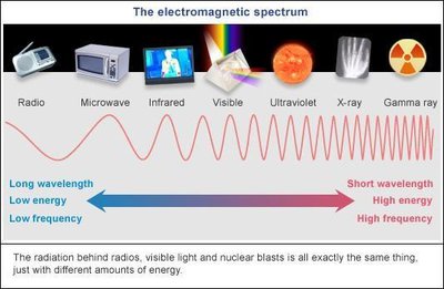
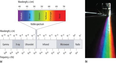
Chromophores and Absorption
Chromophores are functional groups in organic compounds responsible for absorption in the UV/Vis region. Conjugated systems and metal complexes can shift absorption to longer wavelengths and increase intensity.
Common chromophores: Benzene ring, ketones, amines, nitrogen derivatives.
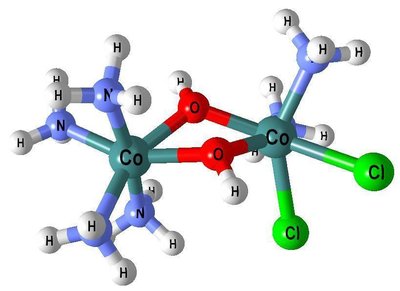
Metal complexes: Transition metal complexes exhibit color due to selective absorption and reflection of visible light.
Instrumentation of UV/Vis Spectroscopy
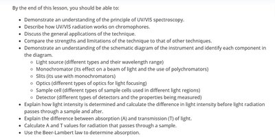
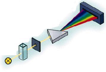
The UV/Vis spectrometer consists of several key components:
Light Source: Deuterium lamp for UV (190–350 nm), quartz halogen/tungsten lamp for visible (350–900 nm).
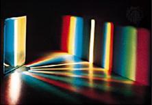
Monochromator: Breaks light into wavelength fractions and isolates the analytical wavelength ().
Slit: Cuts out stray light, allowing only the desired wavelength to pass.
Sample Cell (Cuvette): Quartz for UV, glass/plastic for visible; contains the sample solution.
Detector: Photomultiplier tube or diode array; converts light intensity into electrical signals.
Beer-Lambert Law
The Beer-Lambert Law relates absorbance to concentration:
Absorbance (A):
Transmittance (T):
Beer-Lambert Law:
= molar absorptivity, = pathlength (cm), = concentration (mol/L)
Example: Calculate absorbance for a solution with known concentration and molar absorptivity.
Applications of UV/Vis Spectroscopy
UV/Vis spectroscopy is widely used in chemistry, biochemistry, clinical diagnostics, and industrial applications.
Quantification of active ingredients in formulations
Determination of pKa values
Partition coefficients and solubility studies
Dissolution testing and reaction kinetics monitoring
Strengths and Limitations
Strengths: Easy-to-use, inexpensive, robust, good precision, non-destructive, limited interferences in simple samples.
Limitations: Moderately selective, not suitable for complex samples, limited sensitivity for trace analysis.
Atomic Spectroscopy Techniques
Atomic Absorption Spectroscopy (AAS)
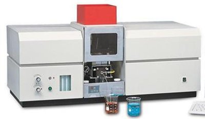
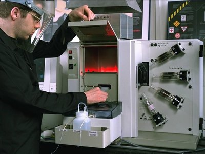
AAS is used to determine the concentration of metallic elements by measuring the absorption of light by atoms in the ground state. A hollow cathode lamp specific to the element provides the excitation energy.
Principle: Atoms absorb light at element-specific wavelengths; absorption is proportional to concentration.
Instrumentation: Hollow cathode lamp, nebulizer, burner/flame, monochromator, detector.
Flame Emission Spectroscopy (FES)
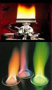
FES quantifies alkali and alkaline earth metals by measuring the emission of light from thermally excited atoms.
Principle: Atoms emit light at specific wavelengths when electrons return to the ground state.
Instrumentation: Burner/flame, nebulizer, monochromator, detector.
Sample Introduction and Nebulization
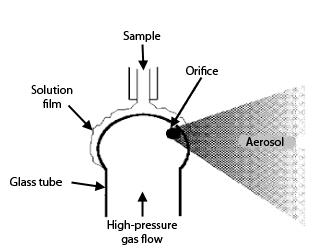
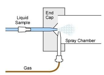
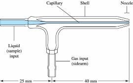
Samples are introduced as aerosols using various types of nebulizers, which convert liquid samples into fine droplets for atomization in the flame.
Pneumatic, Babington, Cross-flow, and Concentric nebulizers are used depending on sample type and analysis requirements.
Interferences in AAS and FES
Several chemical and physical interferences can affect the accuracy of atomic spectroscopy:
Ionisation: High temperatures can ionize atoms, reducing emission/absorption. Solution: Add easily ionizable elements.
Viscosity: Affects nebulizer uptake rate. Solution: Adjust viscosity with ethanol or glycerol.
Anionic Interference: Anions form non-volatile salts with analytes. Solution: Add SrCl2 or LaCl3 to precipitate interfering anions.
Spectral Interference: Overlapping wavelengths from other elements. Solution: Measure at secondary wavelengths.
Applications, Strengths, and Limitations of AAS and FES
Applications: Determination of metals in biological, environmental, and industrial samples.
Strengths: High sensitivity, specificity, limited interferences.
Limitations: Only metallic elements, single-element analysis, destructive (FES).
Inductively Coupled Plasma Optical Emission Spectroscopy (ICP-OES)
Principle and Instrumentation
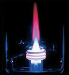
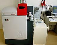
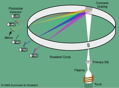
ICP-OES uses an argon plasma to excite atoms and ions, which emit radiation at element-specific wavelengths. A polychromator and diode array detector allow simultaneous multi-element analysis.
Principle: Excited electrons emit radiation; emission intensity is proportional to concentration.
Instrumentation: Nebulizer, plasma torch, polychromator, multiple detectors.
Applications, Strengths, and Limitations of ICP-OES
Applications: Detection of most elements in industrial, environmental, forensic, clinical, and mining samples.
Strengths: High sensitivity, rapid simultaneous analysis, detects most elements including non-metals.
Limitations: Requires skilled operation, expensive instrumentation.
Comparison Table of Spectroscopy Techniques
Technique | Working Principle | Instrumentation | Application | Sample Type/Preparation | Sample Recovery | Strengths | Limitations |
|---|---|---|---|---|---|---|---|
UV-Visible | Absorption of UV/Vis light by molecules | Light source, monochromator, cuvette, detector | Organic/inorganic analysis, drug testing | Translucent solutions | Non-destructive | Easy, inexpensive, robust | Moderate selectivity, limited for complex samples |
AAS | Absorption of light by atoms in ground state | Hollow cathode lamp, nebulizer, burner, detector | Metal analysis in biological/environmental samples | Liquid samples, aerosol | Destructive | High sensitivity, specificity | Single-element, only metals |
FES | Emission of light by thermally excited atoms | Burner, nebulizer, monochromator, detector | Alkali/alkaline earth metal analysis | Liquid samples, aerosol | Destructive | Robust, cheap, selective | Only alkali/alkaline earth metals |
ICP-OES | Emission of light by plasma-excited atoms/ions | Nebulizer, plasma torch, polychromator, detectors | Multi-element analysis in various samples | Liquid samples, aerosol | Destructive | High sensitivity, rapid, multi-element | Expensive, skilled operation |
Additional info: Academic context was added to clarify the principles, instrumentation, and applications of each technique, as well as to expand on the strengths and limitations for exam preparation.
