 Back
BackAdaptive Immunity: Antibody-Mediated Immunity (ANP College Study Notes)
Study Guide - Smart Notes
 Tailored notes based on your materials, expanded with key definitions, examples, and context.
Antibody-Mediated Immunity
Introduction to Antibody-Mediated Immunity
Antibody-mediated immunity is a critical component of the adaptive immune system, primarily involving B cells and antibodies. B cells possess unique receptors that bind to specific antigens, and groups of B cells targeting the same antigen are referred to as clones. When activated, B cells secrete antibodies that bind to the same antigen as their receptors, facilitating antigen destruction.
B cell receptors: Proteins on B cell surfaces that recognize specific antigens.
Antibody: Protein secreted by B cells, also known as immunoglobulin (Ig).
Clone: Group of B cells with identical antigen specificity.
Antibody-mediated immune responses occur in three phases:
B cell clone recognizes its specific antigen and begins secreting antibodies.
Antibody levels in blood rise, leading to antigen destruction.
Memory B cells persist, enabling rapid response upon re-exposure to the antigen.
B Cell Activation, Clonal Selection, and Differentiation
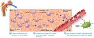
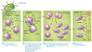
B cells develop and mature in the bone marrow, originating from the lymphoid cell line. Only B cells that do not recognize self-antigens survive, preventing autoimmunity. Mature B cells enter circulation and reside in lymphoid organs, such as the spleen and lymph nodes. Upon encountering their specific antigen, B cells undergo activation, clonal selection, and differentiation.
Autoimmunity: Condition where B cells recognize self-antigens as foreign, producing autoantibodies.
Clonal selection: Process where antigen binds to B cell receptor, activating the B cell.
Cell-Cell Communication: Sensitized B cell presents antigen on class II MHC molecules and interacts with T helper (TH) cells for full activation.
Plasma cells: Differentiated B cells that secrete antibodies.
Memory B cells: Long-lived cells that respond rapidly upon subsequent antigen exposure.
Antibodies and Their Effects
Structure and Classes of Antibodies
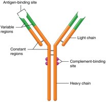
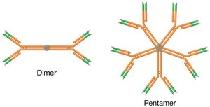
Antibodies are Y-shaped molecules composed of four peptide chains: two heavy (H) and two light (L) chains. Each chain has constant (C) and variable (V) regions. The variable region is responsible for antigen recognition, while the constant region mediates antibody effects. Antibodies can exist as monomers, dimers, or pentamers, depending on their class.
Constant region (C): Similar among antibody classes; responsible for antibody functions.
Variable region (V): Unique sequence for antigen binding.
Monomer: Single antibody unit.
Dimer: Two antibody units.
Pentamer: Five antibody units.
Antibody Classes and Functions
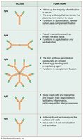
There are five basic classes of antibodies, each with distinct structures and functions. The classes are designated as IgG, IgA, IgM, IgE, and IgD, remembered by the mnemonic GAMED.
IgG: Most prevalent; crosses placenta; single subunit.
IgA: Dimer; found in secretions (tears, saliva, sweat, breast milk).
IgM: Pentamer; first antibody secreted; potent agglutinating agent.
IgE: Single subunit; binds to allergens and parasitic antigens; triggers inflammation.
IgD: Single subunit; acts as B cell receptor; not secreted in significant amounts.
Functions of Secreted Antibodies
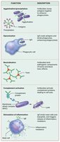
Antibodies bind antigens, leading to several effects on pathogens:
Agglutination: Antibodies cross-link cells, forming clumps for easier phagocytosis.
Precipitation: Antibodies bind soluble antigens, decreasing their solubility.
Opsonization: Antibodies (especially IgG) coat pathogens, enhancing phagocytosis.
Neutralization: Antibodies block toxins and viral proteins from interacting with cells.
Complement activation: IgM and IgG activate complement proteins, aiding pathogen destruction.
Stimulation of inflammation: IgE triggers release of inflammatory mediators.
Immunological Memory
Primary and Secondary Immune Responses
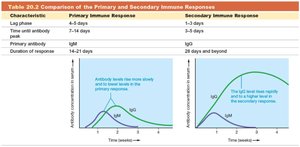
Memory B cells enable rapid and efficient responses upon re-exposure to antigens. The primary immune response occurs upon first exposure, with a lag phase of 4–5 days and peak antibody levels at 7–14 days. The secondary immune response is faster (1–3 days lag), with higher and more effective antibody levels, primarily involving IgG.
Primary response: Slow, mainly IgM, lag phase, moderate antibody levels.
Secondary response: Rapid, mainly IgG, higher affinity, larger antibody peak.
Characteristic | Primary Immune Response | Secondary Immune Response |
|---|---|---|
Lag phase | 4–5 days | 1–3 days |
Antibody peak | 7–14 days | 3–5 days |
Primary antibody | IgM | IgG |
Duration | 14–21 days | 28 days and beyond |
Vaccination and Types of Immunity
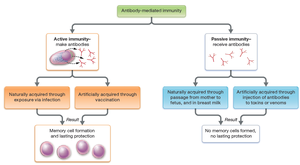
Vaccination exposes individuals to antigens, eliciting a primary immune response and generating memory cells. Upon subsequent exposure, the secondary response minimizes symptoms. There are two types of antibody-mediated immunity: active and passive.
Active immunity: Body actively responds to antigen; acquired naturally (infection) or artificially (vaccination); produces memory cells; long-lasting.
Passive immunity: Preformed antibodies are transferred; acquired naturally (mother to fetus) or artificially (injection); no memory cells; short-lived.
Pseudoscience Exposed: Vaccines and Autism
Debunking the Vaccine-Autism Myth
Despite widespread claims, scientific research has found no link between vaccines and autism. The concern originated from the timing of childhood vaccinations and the use of thimerosal, a mercury-containing preservative. However, thimerosal contains ethylmercury, not methylmercury (the neurotoxic form). Studies have consistently shown no association between thimerosal and autism, and removal of thimerosal from vaccines did not decrease autism rates. Continued belief in this myth has led to reduced vaccination rates and increased cases of preventable diseases, posing significant public health risks.
Thimerosal: Ethylmercury-containing preservative; not linked to autism.
Public health impact: Decreased vaccination rates increase disease incidence.
