 Back
BackAnatomy & Physiology Exam 1 Study Guide: Foundations, Chemistry, Cells, and Integument
Study Guide - Smart Notes
 Tailored notes based on your materials, expanded with key definitions, examples, and context.
Chapter 1: Introduction to Anatomy and Physiology
Characteristics of Life
Living organisms share several defining characteristics that distinguish them from non-living matter.
Organization: Living things exhibit a complex but ordered structure.
Metabolism: The sum of all chemical reactions occurring in the body, including catabolism (breaking down molecules) and anabolism (building molecules).
Responsiveness: Ability to sense and respond to stimuli.
Growth: Increase in size and number of cells.
Development: Changes in form and function during an organism's life cycle.
Reproduction: Production of new organisms and cells.
Regulation: Ability to maintain internal stability (homeostasis).
Adaptation: Evolutionary changes that enhance survival.
Conditions That Support Life & Essential Nutrients
Life is supported by specific environmental conditions and nutrients:
Water: Essential for metabolic reactions and transport.
Nutrients: Carbohydrates, proteins, lipids, vitamins, and minerals are required for energy, growth, and repair.
Oxygen: Required for cellular respiration.
Stable Temperature & Pressure: Necessary for proper metabolic function.
Major Fluid Compartments
The body’s fluids are distributed in distinct compartments:
Intracellular Fluid (ICF): Fluid within cells; high in potassium (K+).
Extracellular Fluid (ECF): Fluid outside cells; high in sodium (Na+).
Interstitial Fluid: Surrounds tissue cells.
Plasma: Fluid component of blood.
Other: Lymph, cerebrospinal fluid, etc.
Homeostasis
Homeostasis is the maintenance of a stable internal environment despite external changes. It is essential for survival and function.
Regulated by feedback mechanisms that detect and respond to deviations from set points.
Homeostatic Feedback Loops
Feedback loops are the primary mechanisms for maintaining homeostasis. They involve:
Receptor: Detects changes in the environment.
Control Center: Processes information and determines response.
Effector: Carries out the response to restore balance.
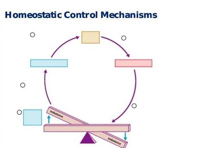
Negative Feedback: Reduces the effect of the stimulus, restoring balance (e.g., body temperature regulation).
Positive Feedback: Enhances the effect of the stimulus (e.g., blood clotting, labor contractions).
Chapter 2: The Chemistry of Life
Kinetic and Potential Energy
Kinetic energy is energy of motion, while potential energy is stored energy due to position or structure.
Example: Chemical bonds store potential energy; when bonds break, energy is released as kinetic energy.
Atomic Structure & Subatomic Particles
Atoms consist of a nucleus (protons and neutrons) and electrons orbiting the nucleus.
Protons (p+): Positively charged, in nucleus.
Neutrons (n0): Neutral, in nucleus.
Electrons (e-): Negatively charged, orbit nucleus.
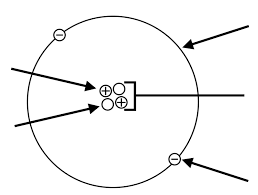
Atomic Variation: Ions vs Isotopes
Ions: Atoms with a net charge due to loss or gain of electrons (e.g., Na+, Cl-).
Isotopes: Atoms with the same number of protons but different numbers of neutrons (e.g., Carbon-12 vs Carbon-14).
Chemical Bonds
Covalent Bonds: Atoms share electrons (e.g., H2O).
Ionic Bonds: Transfer of electrons from one atom to another, resulting in charged ions (e.g., NaCl).
Water and Hydrogen Bonding
Water is a polar molecule, allowing it to form hydrogen bonds, which are weak attractions between the hydrogen atom of one molecule and an electronegative atom of another.
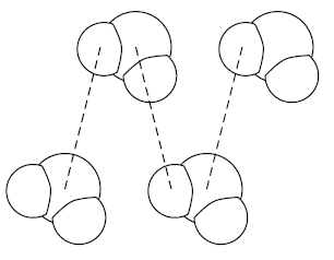
Hydrogen bonds give water its unique properties: high heat capacity, cohesion, adhesion, and solvent abilities.
pH: Acids vs Bases
pH measures the concentration of hydrogen ions (H+) in a solution.
Acids: Release H+ (pH < 7).
Bases: Accept H+ (pH > 7).
Carbonic Acid/Bicarbonate Buffering Mechanism
This buffer system helps maintain blood pH:
$\mathrm{CO_2 + H_2O \leftrightarrow H_2CO_3 \leftrightarrow H^+ + HCO_3^-}$
Organic Molecules: General Characteristics
Organic molecules contain carbon and hydrogen, and are often large and complex.
The Four Classes of Organic Molecules
Lipids: Nonpolar molecules used for energy storage, insulation, and cell membranes (e.g., triglycerides, phospholipids).
Proteins: Polymers of amino acids; serve as enzymes, structural components, and signaling molecules.
Carbohydrates: Sugars and starches; primary energy source for cells.
Nucleic Acids: DNA and RNA; store and transmit genetic information.
Chapter 3: The Cell
Cellular Membranes
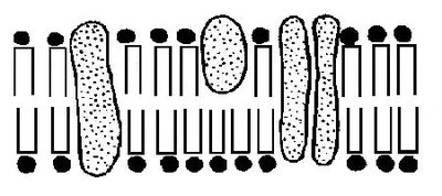
The plasma membrane is a selectively permeable barrier composed of a phospholipid bilayer with embedded proteins, cholesterol, and carbohydrates.
Phospholipids: Form the basic structure; hydrophilic heads and hydrophobic tails.
Proteins: Serve as channels, receptors, enzymes, and anchors.
Carbohydrates: Involved in cell recognition and signaling.
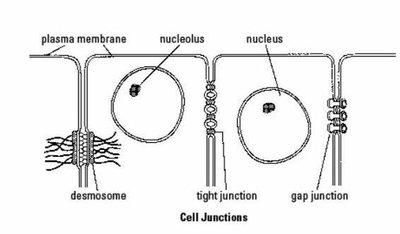
Cellular Junctions
Specialized structures connect adjacent cells:
Tight Junctions: Seal cells together, preventing passage of substances between them.
Desmosomes: Anchor cells together, providing mechanical strength.
Gap Junctions: Allow communication and passage of ions/small molecules between cells.
Membrane Specializations
Microvilli: Increase surface area for absorption (e.g., intestinal cells).
Cilia: Move substances across cell surfaces (e.g., respiratory tract).
Flagella: Enable cell movement (e.g., sperm cells).
Membrane Transport
Movement of substances across the membrane depends on membrane composition and solute properties.
Passive Transport: Does not require energy.
Simple Diffusion: Movement of small, nonpolar molecules down their concentration gradient.
Facilitated Diffusion: Movement via membrane proteins (channels/carriers).
Osmosis: Diffusion of water across a semipermeable membrane.
Osmotic Pressure: Pressure required to stop osmosis.
Hydrostatic Pressure: Pressure exerted by a fluid.
Osmolarity: Total solute concentration of a solution.
Tonicity: Effect of a solution on cell volume (hypertonic, isotonic, hypotonic).
Active Transport: Requires energy (ATP).
Primary Active Transport: Direct use of ATP (e.g., sodium/potassium pump).
Secondary Active Transport: Uses energy from ion gradients.
Vesicular/Bulk Transport: Movement of large particles via vesicles (endocytosis, exocytosis).
Protein Synthesis
Protein synthesis involves two main processes:
Transcription: DNA is transcribed into messenger RNA (mRNA) in the nucleus.
Translation: mRNA is translated into a protein at the ribosome.
DNA Bases: Adenine (A), Thymine (T), Cytosine (C), Guanine (G)
RNA Bases: Adenine (A), Uracil (U), Cytosine (C), Guanine (G)
Base Pairing: A-T (DNA), A-U (RNA), C-G
Cell Cycle
Interphase: Includes G1 (growth), S (DNA synthesis), G2 (preparation for division), and G0 (resting phase).
Mitosis: Division of the nucleus into two identical daughter cells.
Cellular Death and Adaptation
Necrosis: Unplanned cell death due to injury.
Apoptosis: Programmed cell death; normal and controlled.
Autophagy: Cell digests its own components for recycling.
Chapter 5: The Integumentary System
Anatomy of the Skin
The skin consists of three main layers:
Epidermis: Outermost, avascular layer composed of stratified squamous epithelium.
Dermis: Middle, vascular layer containing connective tissue, nerves, and blood vessels.
Hypodermis (Subcutaneous): Deepest layer, primarily adipose tissue for insulation and energy storage.
Tissues & Cell Types in the Skin
Melanocytes: Produce melanin pigment for UV protection.
Keratinocytes: Main cell type; produce keratin for waterproofing and protection.
Dendritic (Langerhans) Cells: Immune defense.
Merkel Cells: Sensory receptors for touch.
Fibroblasts: Produce collagen and extracellular matrix in the dermis.
Mast Cells: Involved in inflammation and allergic responses.
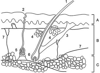
Appendages of the Skin
Glands:
Sebaceous: Secrete sebum (oil) for lubrication.
Sudoriferous: Sweat glands for thermoregulation.
Ceruminous: Produce earwax.
Apocrine: Scent glands in specific areas.
Hair: Protection, sensation, and insulation.
Nails: Protect fingertips and enhance sensation.
Vitamin D Synthesis and Function
Skin plays a critical role in vitamin D synthesis, which is essential for calcium absorption and bone health.
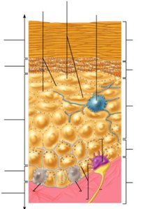
Melanin Production, Dispersal, and Function
Melanin is produced by melanocytes and dispersed to keratinocytes, providing pigmentation and protection from UV radiation.
Biological Protection
Epidermal Defenses: Acid mantle, defensins, dermcidin, and Langerhans cells provide antimicrobial and immune protection.
Dermal Defenses: Mast cells and leukocytes participate in immune responses and inflammation.
Physical Protection
Epidermal: Keratin, lipid barrier, and cell junctions prevent water loss and entry of pathogens.
Dermal: Collagen and elastic fibers provide strength and flexibility.
Thermoregulation
The skin regulates body temperature through sweat production and blood flow adjustments.
Avenues for Heat Loss: Radiation, conduction, convection, and evaporation.
Wound Response and Healing
Inflammatory Response: Characterized by redness, heat, swelling, and pain; mast cells release histamine.
Superficial Wound Repair: Involves regeneration of epidermal cells.
Deep Wound Repair: Involves inflammation, proliferation, and remodeling phases.
Burns: Extent and Rule of Nines
First Degree: Affects only the epidermis; redness and pain.
Second Degree: Involves epidermis and part of dermis; blisters.
Third Degree: Destroys entire skin layer; risk of fluid loss and infection.
Rule of Nines: Used to estimate the percentage of body surface area affected by burns.
Skin Tumors
Basal Cell Carcinoma: Most common, least dangerous; arises from basal cells.
Squamous Cell Carcinoma: Arises from keratinocytes; can metastasize.
Melanoma: Most dangerous; arises from melanocytes; high metastatic potential.
