 Back
BackAnatomy & Physiology Exam Study Guidance
Study Guide - Smart Notes
 Tailored notes based on your materials, expanded with key definitions, examples, and context.
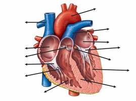
Q1. Label the parts of the heart.
Background
Topic: Heart Anatomy
This question tests your knowledge of the internal structure of the heart, including chambers, valves, and major vessels. Understanding heart anatomy is foundational for learning about cardiovascular physiology and pathology.
Key Terms:
Atria: The upper chambers of the heart (right and left atrium).
Ventricles: The lower chambers (right and left ventricle).
Valves: Structures that ensure one-way blood flow (tricuspid, bicuspid/mitral, pulmonary, aortic).
Major vessels: Superior/inferior vena cava, pulmonary arteries/veins, aorta.
Step-by-Step Guidance
Identify the four chambers: two atria (upper) and two ventricles (lower).
Locate the major vessels entering and leaving the heart: superior/inferior vena cava, pulmonary arteries, pulmonary veins, and aorta.
Find the valves between chambers and vessels: tricuspid (right atrium to right ventricle), pulmonary (right ventricle to pulmonary artery), bicuspid/mitral (left atrium to left ventricle), and aortic (left ventricle to aorta).
Observe the direction of blood flow through the heart, noting which structures are involved at each step.
Try solving on your own before revealing the answer!
Final Answer:
The labeled parts of the heart include: right atrium, right ventricle, left atrium, left ventricle, tricuspid valve, pulmonary valve, bicuspid (mitral) valve, aortic valve, superior vena cava, inferior vena cava, pulmonary arteries, pulmonary veins, and aorta.
These structures are essential for the heart's function as a pump, ensuring blood flows in the correct direction through the pulmonary and systemic circuits.
Q2. What is the blood flow through the heart?
Background
Topic: Heart Physiology
This question tests your understanding of the sequence of blood flow through the heart's chambers, valves, and vessels. Knowing this pathway is crucial for understanding oxygenation and circulation.
Key Terms:
Pulmonary circuit: Pathway from the right side of the heart to the lungs and back.
Systemic circuit: Pathway from the left side of the heart to the body and back.
Valves: Ensure unidirectional flow.
Step-by-Step Guidance
Start with deoxygenated blood entering the right atrium via the superior and inferior vena cava.
Blood passes through the tricuspid valve into the right ventricle.
From the right ventricle, blood is pumped through the pulmonary valve into the pulmonary arteries, which carry it to the lungs.
Oxygenated blood returns from the lungs via the pulmonary veins to the left atrium.
Try solving on your own before revealing the answer!
Final Answer:
Blood flow through the heart: Right atrium → tricuspid valve → right ventricle → pulmonary valve → pulmonary arteries → lungs → pulmonary veins → left atrium → bicuspid (mitral) valve → left ventricle → aortic valve → aorta → body.
This sequence ensures proper oxygenation and circulation throughout the body.
Q3. Describe the difference between the Intrinsic and Extrinsic conduction system of the heart.
Background
Topic: Cardiac Conduction System
This question tests your understanding of how the heart's electrical activity is generated and regulated, both internally (intrinsic) and externally (extrinsic).
Key Terms:
Intrinsic conduction system: Specialized cardiac cells (SA node, AV node, bundle of His, Purkinje fibers) that generate and propagate electrical impulses.
Extrinsic conduction system: External influences (autonomic nervous system, hormones) that modulate heart rate and force.
Step-by-Step Guidance
Define the intrinsic conduction system and list its main components.
Explain how the intrinsic system initiates and coordinates the heartbeat.
Describe the extrinsic conduction system and its role in modifying heart activity.
Compare the two systems in terms of their origin and function.
Try solving on your own before revealing the answer!
Final Answer:
The intrinsic conduction system consists of specialized cardiac cells that generate and propagate electrical impulses, setting the basic rhythm of the heart. The extrinsic conduction system involves external factors (such as the autonomic nervous system and hormones) that adjust heart rate and force in response to physiological needs.
Intrinsic = internal pacemaker; Extrinsic = external modulation.
Q4. What are the lymphoid cells (T lymphocytes and B lymphocytes)? Where do they both originate? Where do they mature?
Background
Topic: Lymphatic System and Immunity
This question tests your knowledge of the cells responsible for adaptive immunity and their development.
Key Terms:
T lymphocytes (T cells): Cells involved in cell-mediated immunity.
B lymphocytes (B cells): Cells responsible for humoral immunity (antibody production).
Origin: Both types originate from hematopoietic stem cells in the bone marrow.
Maturation: T cells mature in the thymus; B cells mature in the bone marrow.
Step-by-Step Guidance
Identify the two main types of lymphoid cells and their roles in immunity.
Describe where both T and B lymphocytes originate.
Explain the maturation process for each cell type and the organs involved.
Relate the maturation sites to their function in the immune system.
Try solving on your own before revealing the answer!
Final Answer:
T lymphocytes and B lymphocytes both originate from stem cells in the bone marrow. T cells mature in the thymus, while B cells mature in the bone marrow. T cells are involved in cell-mediated immunity; B cells produce antibodies for humoral immunity.
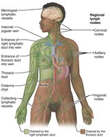
Q5. What is the function of the spleen?
Background
Topic: Lymphatic System – Spleen
This question tests your understanding of the spleen's role in immunity and blood filtration.
Key Terms:
Spleen: Largest lymphoid organ, located in the upper left abdomen.
White pulp: Immune function (lymphocyte proliferation).
Red pulp: Blood filtration, removal of old RBCs, storage of platelets.
Step-by-Step Guidance
Identify the anatomical location and structure of the spleen.
Describe the two main regions: white pulp and red pulp.
Explain the immune functions of the white pulp.
Discuss the blood filtration and storage functions of the red pulp.
Try solving on your own before revealing the answer!
Final Answer:
The spleen filters blood, removes old or damaged red blood cells, stores platelets, and provides an immune response through its white pulp. It is essential for both blood maintenance and immune defense.
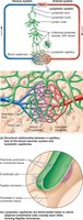
Q6. What is the structure of a lymph node?
Background
Topic: Lymphatic System – Lymph Node Anatomy
This question tests your knowledge of the internal structure and function of lymph nodes, which are key sites for immune cell activation.
Key Terms:
Cortex: Contains lymphoid follicles and germinal centers.
Medulla: Contains medullary cords and sinuses.
Capsule: Outer covering.
Afferent/efferent vessels: Entry and exit points for lymph.
Step-by-Step Guidance
Identify the main regions: cortex and medulla.
Describe the function of lymphoid follicles and germinal centers in the cortex.
Explain the role of medullary cords and sinuses in the medulla.
Locate the capsule and the afferent/efferent lymphatic vessels.
Try solving on your own before revealing the answer!
Final Answer:
A lymph node consists of an outer cortex (with follicles and germinal centers), an inner medulla (with medullary cords and sinuses), a capsule, and afferent/efferent lymphatic vessels. It filters lymph and houses immune cells.
