 Back
BackAnatomy & Physiology II: The Reproductive System (Chapter 27) Study Notes
Study Guide - Smart Notes
 Tailored notes based on your materials, expanded with key definitions, examples, and context.
Reproductive System Overview
Introduction to Human Reproduction
The human reproductive system is responsible for producing, storing, and delivering gametes (sex cells) and for the secretion of sex hormones. It ensures the continuation of the species through sexual reproduction.
Gametes: Specialized sex cells (sperm in males, oocyte in females), each with n = 23 chromosomes.
Gonads: Organs that produce gametes and sex hormones (testes in males, ovaries in females).
Androgens: Sex hormones such as testosterone (males) and estrogen/progesterone (females).
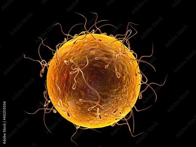
Male Reproductive System
Anatomy of the Male Reproductive System
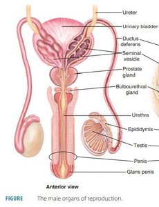
The male reproductive system includes both internal and external structures that function in the production, maturation, and delivery of sperm.
Testes: Produce sperm and testosterone.
Scrotum: Sac that holds the testes and regulates their temperature.
Epididymis: Site of sperm maturation and storage.
Ductus (vas) deferens: Transports sperm from the epididymis to the urethra.
Accessory glands: Seminal vesicles, prostate gland, and bulbourethral glands contribute fluids to semen.
Penis: Organ for copulation and sperm delivery.
Sperm Transport Pathway
Sperm are produced in the testes and transported through a series of ducts and glands before ejaculation.
Seminiferous tubules → rete testis
Epididymis (storage and maturation)
Ductus (vas) deferens (within spermatic cord, through inguinal canal)
Seminal vesicle (adds fructose-rich fluid)
Prostate gland (adds alkaline, milky fluid)
Ejaculatory duct
Bulbourethral gland (adds lubricating, cleansing fluid)
Penis (expulsion of semen)
Testes and Seminiferous Tubules
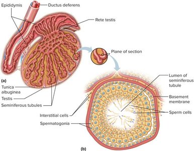
The testes are covered by two protective layers and contain seminiferous tubules where sperm develop.
Tunica albuginea: Dense connective tissue covering.
Tunica vaginalis: Serous membrane.
Muscles: Dartos (scrotal septum) and cremaster (temperature regulation).
Spermatogenesis
Spermatogenesis is the process of sperm cell development, taking approximately 64 days and involving both mitosis and meiosis.
Spermatogonia (2n): Stem cells divide by mitosis.
Primary spermatocytes (2n): Undergo meiosis I to form secondary spermatocytes (n).
Secondary spermatocytes (n): Undergo meiosis II to form spermatids (n).
Spermiogenesis: Spermatids mature into spermatozoa (sperm).
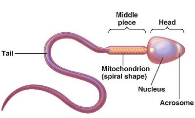
Sperm Structure:
Head: Contains nucleus (DNA) and acrosome (enzymes for egg penetration).
Midpiece: Packed with mitochondria for ATP production.
Tail (flagellum): Propels sperm.
Semen and Accessory Glands
Semen is the fluid ejaculated during orgasm, containing sperm and secretions from accessory glands.
Seminal vesicle: Produces 60% of semen volume, rich in fructose.
Prostate gland: Adds alkaline, milky fluid to neutralize vaginal acidity.
Bulbourethral (Cowper’s) gland: Secretes lubricating, cleansing fluid (pre-ejaculate).
Penis Structure and Erection
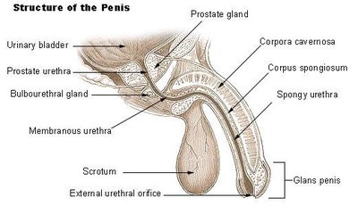
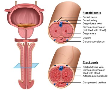
The penis contains erectile tissue that fills with blood during sexual arousal, resulting in an erection.
Corpora cavernosa: Two dorsal columns of erectile tissue.
Corpus spongiosum: Surrounds the urethra and forms the glans penis.
Erection: Mediated by nitric oxide (NO), which relaxes smooth muscle and increases blood flow.
Hormonal Regulation in Males
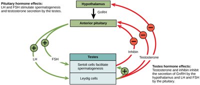
Testosterone production and spermatogenesis are regulated by the hypothalamic-pituitary-gonadal axis.
Leydig cells: Produce testosterone in response to LH.
Sertoli cells: Support spermatogenesis, produce inhibin.
GnRH (Gonadotropin-releasing hormone): From hypothalamus, stimulates FSH and LH release from anterior pituitary.
Negative feedback: Testosterone and inhibin inhibit GnRH, FSH, and LH secretion.
Female Reproductive System
External Female Genitalia
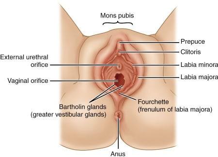
The external female genitalia, collectively called the vulva, protect the internal reproductive organs and facilitate sexual intercourse.
Mons pubis: Fatty area over the pubic bone.
Labia majora and minora: Folds of skin that protect the vaginal and urethral openings.
Clitoris: Erectile tissue, homologous to the glans penis.
Hymen: Thin membrane at the vaginal entrance.
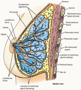
Breasts and Mammary Glands
The breasts are accessory organs of the female reproductive system, primarily functioning in lactation.
Areola: Pigmented area around the nipple, contains glands for lubrication.
Mammary glands: Modified sweat glands that produce milk.
Lactiferous ducts and sinuses: Transport and store milk.
Myoepithelial cells: Contract to expel milk.
Suspensory ligaments: Support the breast tissue.
Internal Female Reproductive Organs
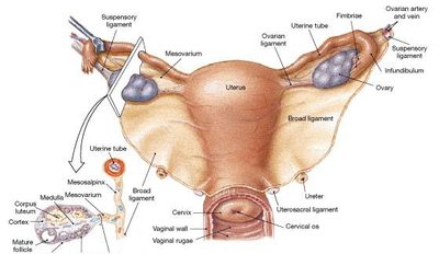
The internal organs include the ovaries, uterine tubes, uterus, and vagina, each with specialized functions in reproduction.
Ovaries: Produce oocytes and sex hormones.
Uterine (fallopian) tubes: Transport oocytes from ovary to uterus; site of fertilization.
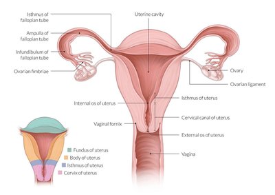
Uterus: Site of implantation and fetal development; consists of fundus, body, and cervix.
Vagina: Muscular canal for intercourse, childbirth, and menstrual flow.
Uterine Tubes (Fallopian Tubes)
The uterine tubes are the conduit for oocytes and the typical site of fertilization.
Isthmus: Narrow region closest to the uterus.
Infundibulum: Funnel-shaped region with fimbriae that sweep oocytes into the tube.
Ampulla: Widest region, common site of fertilization.
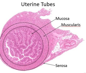
Layers: Mucosa (ciliated), muscularis (smooth muscle), serosa (outer layer).
Ovarian and Menstrual Cycles
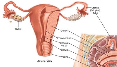
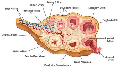
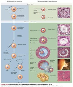
The ovarian cycle (oogenesis and folliculogenesis) and the menstrual cycle are coordinated to ensure successful reproduction.
Oogenesis: Formation of oocytes begins before birth and continues through reproductive years.
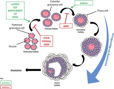
Folliculogenesis: Development of ovarian follicles, from primordial to mature (Graafian) follicle.
Ovulation: Release of a secondary oocyte, triggered by LH surge.
Corpus luteum: Secretes progesterone; degenerates if no pregnancy occurs.
Hormonal Regulation in Females
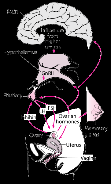
Female reproductive function is regulated by the hypothalamic-pituitary-ovarian axis.
GnRH: Stimulates FSH and LH release from the anterior pituitary.
FSH: Promotes follicular growth and estrogen production.
LH: Triggers ovulation and corpus luteum formation.
Estrogen and progesterone: Regulate the menstrual cycle and prepare the endometrium for implantation.
Uterine (Menstrual) Cycle
The menstrual cycle prepares the endometrium for possible implantation and is divided into three phases:
Menses: Shedding of the endometrial lining (days 1–7).
Proliferative phase: Endometrial regeneration and thickening (days 7–14), ends with ovulation.
Secretory phase: Endometrial preparation for implantation, driven by progesterone (days 15–28).
Menarche: First menstruation after puberty.
Corpus luteum: Maintains endometrium; degenerates if no pregnancy, leading to menses.
Cell Division: Mitosis and Meiosis
Cell Cycle and Mitosis
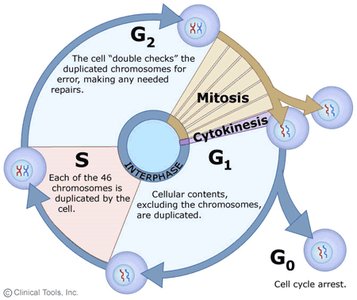
Somatic cells divide by mitosis, producing genetically identical diploid cells (2n = 46 chromosomes).
Interphase: G1 (growth), S (DNA synthesis), G2 (preparation for division).
Mitosis: Prophase, metaphase, anaphase, telophase.
Cytokinesis: Division of cytoplasm.
Meiosis
Meiosis produces gametes with half the chromosome number (n = 23), introducing genetic diversity.
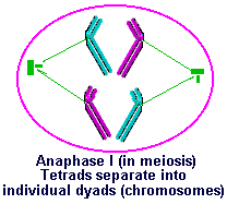
Meiosis I: Homologous chromosomes separate (tetrads split in anaphase I).
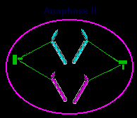
Meiosis II: Sister chromatids separate (dyads split in anaphase II).
Crossing over: Occurs in prophase I, increasing genetic variation.
Embryonic Development and Sexual Differentiation
Development of Gonads
Primordial gonads develop early in embryogenesis and differentiate into testes or ovaries based on genetic and hormonal signals.
SRY gene: Presence triggers male development (testes, testosterone production).
Absence of SRY: Default development is female.
Müllerian ducts: Develop into female reproductive tract unless suppressed by male hormones.
Wolffian ducts: Develop into male reproductive tract under influence of testosterone.
Puberty and Sexual Maturation
Puberty marks the onset of sexual maturity, with increased hormone production and development of secondary sexual characteristics.
LH and FSH: Levels rise, stimulating gonadal activity.
Secondary sexual characteristics: Develop under influence of sex hormones (e.g., breast development, body hair, voice changes).
Summary Table: Comparison of Male and Female Gametogenesis
Feature | Spermatogenesis (Male) | Oogenesis (Female) |
|---|---|---|
Location | Seminiferous tubules of testes | Ovarian cortex |
Timing | Puberty to old age | Begins before birth, resumes at puberty, ends at menopause |
Number of gametes per cycle | Millions per day | One (usually) per month |
Meiotic divisions | Continuous | Arrested at prophase I until puberty, then at metaphase II until fertilization |
Final products | 4 sperm per primary spermatocyte | 1 ovum + 2-3 polar bodies per primary oocyte |
