 Back
Anatomy & Physiology Study Guide: Nervous and Muscle Tissue
Study Guide - Smart Notes
 Tailored notes based on your materials, expanded with key definitions, examples, and context.
Nervous System: Structure and Function
Overview of the Nervous System
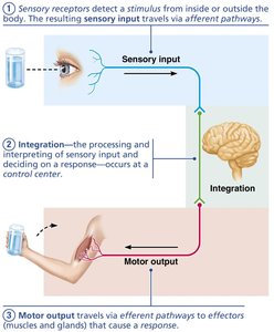
The nervous system is the master controlling and communicating system of the body. It enables rapid and specific communication between cells via electrical and chemical signals, resulting in almost immediate responses.
Sensory Input: Information gathered by sensory receptors about internal and external changes.
Integration: Processing and interpretation of sensory input to decide on a response.
Motor Output: Activation of effectors (muscles and glands) to produce a response.
Divisions of the Nervous System
The nervous system is divided into two principal parts:
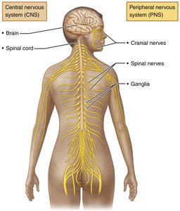
Central Nervous System (CNS): Consists of the brain and spinal cord. It serves as the integration and control center, interpreting sensory input and dictating motor output.
Peripheral Nervous System (PNS): Comprises nerves that extend from the brain and spinal cord, including cranial nerves, spinal nerves, and ganglia. The PNS connects the CNS to the rest of the body.
Functional Divisions of the PNS
Sensory (Afferent) Division: Somatic sensory fibers convey impulses from skin, skeletal muscles, and joints to the CNS; visceral sensory fibers convey impulses from visceral organs.
Motor (Efferent) Division: Transmits impulses from CNS to effectors. Includes the somatic nervous system (voluntary control of skeletal muscles) and the autonomic nervous system (involuntary control of smooth muscle, cardiac muscle, and glands).
Nervous Tissue: Cellular Components
Neuroglia (Glial Cells) of the CNS
Nervous tissue consists of two principal cell types: neuroglia (supporting cells) and neurons (excitable cells).
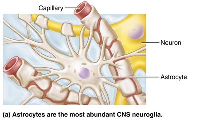
Astrocytes: Most abundant glial cells; support neurons, regulate exchanges between capillaries and neurons, guide migration of young neurons, and control the chemical environment.
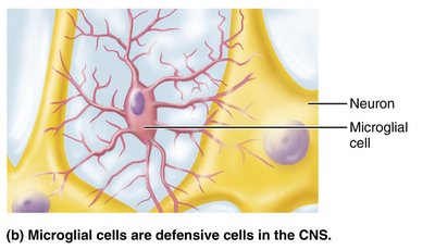
Microglial Cells: Defensive cells that monitor neurons and can transform to phagocytize microorganisms and debris.
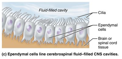
Ependymal Cells: Line cerebrospinal fluid-filled cavities; may be ciliated to help circulate CSF.
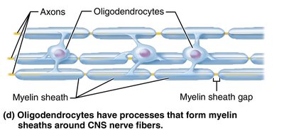
Oligodendrocytes: Form myelin sheaths around CNS nerve fibers, insulating and increasing the speed of impulse transmission.
Neuroglia of the PNS
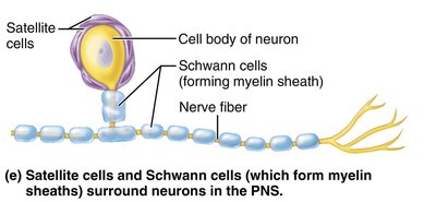
Satellite Cells: Surround neuron cell bodies in the PNS; function similarly to astrocytes.
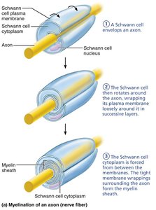
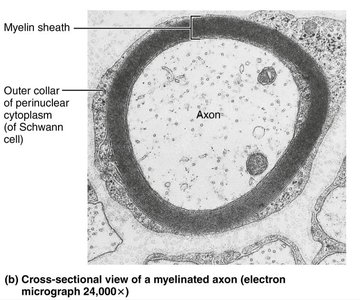
Schwann Cells: Surround all peripheral nerve fibers and form myelin sheaths; vital for regeneration of damaged peripheral nerve fibers.
Neurons: Structure and Function
Neurons are the structural units of the nervous system, specialized for conducting impulses. They exhibit extreme longevity, are mostly amitotic, and have a high metabolic rate requiring continuous oxygen and glucose.
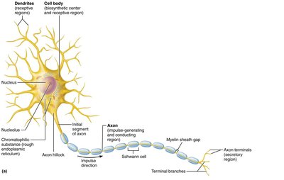
Cell Body (Soma): Contains the nucleus and organelles.
Dendrites: Receptive regions that convey incoming messages toward the cell body.
Axon: Conducting region that generates and transmits nerve impulses away from the cell body to axon terminals, which release neurotransmitters.
Myelination of Axons
Myelin is a white, fatty substance that coats many axons, providing electrical insulation and increasing the speed of impulse transmission. Myelination in the PNS is performed by Schwann cells, while in the CNS it is performed by oligodendrocytes.
Myelin Sheath Gaps (Nodes of Ranvier): Gaps between adjacent Schwann cells where axon collaterals can emerge.
Nonmyelinated Fibers: Thin fibers not wrapped in myelin; conduct impulses more slowly.
Neuronal Physiology: Membrane Potential and Ion Channels
Resting Membrane Potential
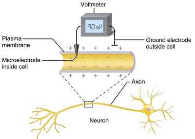
Neurons have a resting membrane potential, typically around −70 mV, with the cytoplasmic side negatively charged relative to the outside. This potential is maintained by differences in ionic composition and selective permeability of the plasma membrane.
Voltage: Potential energy generated by separated charge.
Current: Flow of electrical charge (ions) between two points.
Resistance: Hindrance to charge flow.
Membrane Ion Channels
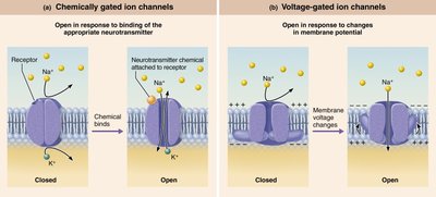
Ion channels are large proteins in the plasma membrane that allow specific ions to pass through. There are two main types:
Leakage (Nongated) Channels: Always open.
Gated Channels: Open and close in response to specific stimuli.
Chemically Gated Channels: Open with binding of a specific chemical (e.g., neurotransmitter).
Voltage-Gated Channels: Open and close in response to changes in membrane potential.
Mechanically Gated Channels: Open and close in response to physical deformation.
Muscle Tissue: Types and Structure
Types of Muscle Tissue
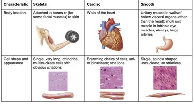
Muscle tissue makes up nearly half of the body’s mass and can transform chemical energy (ATP) into mechanical energy. There are three types:
Skeletal Muscle: Attached to bones; voluntary control; striated.
Cardiac Muscle: Found in the heart; involuntary control; striated with intercalated discs.
Smooth Muscle: Found in walls of hollow organs; involuntary control; non-striated.
Structure of Skeletal Muscle
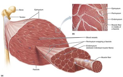
Skeletal muscle is organized into bundles covered by connective tissue:
Epimysium: Surrounds entire muscle.
Perimysium: Surrounds fascicles (groups of muscle fibers).
Endomysium: Surrounds individual muscle fibers.
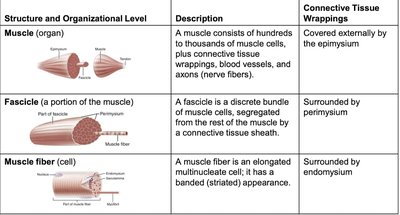
Structure and Organizational Level | Description | Connective Tissue Wrappings |
|---|---|---|
Muscle (organ) | Hundreds to thousands of muscle cells, plus connective tissue, blood vessels, and nerves | Epimysium |
Fascicle | Discrete bundle of muscle cells | Perimysium |
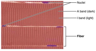
Muscle fiber (cell) | Elongated multinucleate cell with striated appearance | Endomysium |
Skeletal Muscle Fiber Structure
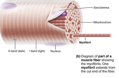
Skeletal muscle fibers are long, cylindrical cells with multiple nuclei. Specialized structures include:
Sarcolemma: Plasma membrane of muscle fiber.
Sarcoplasm: Cytoplasm of muscle fiber, rich in glycogen and myoglobin.
Myofibrils: Densely packed rodlike elements accounting for ~80% of cell volume.
Myofibril Structure: Sarcomeres and Striations
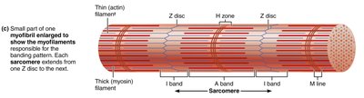
Myofibrils contain repeating units called sarcomeres, the smallest contractile unit of muscle fiber. Striations are formed by alternating dark (A bands) and light (I bands) regions.
A Band: Dark region; contains H zone and M line.
I Band: Light region; contains Z disc.
Myofilaments: Thick and Thin Filaments
Myofilaments are arranged within the sarcomere:
Actin (Thin) Filaments: Extend across I band and partway in A band; anchored to Z discs; contain regulatory proteins tropomyosin and troponin.
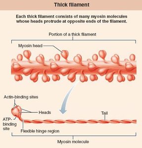
Myosin (Thick) Filaments: Extend length of A band; connected at M line; each myosin molecule has heads for binding actin and ATP.
Summary Table: Muscle Tissue Types
Characteristic | Skeletal | Cardiac | Smooth |
|---|---|---|---|
Body location | Attached to bones or skin | Walls of the heart | Walls of hollow organs (except heart) |
Cell shape and appearance | Long, cylindrical, multinucleate, striated | Branching chains, uni- or binucleate, striated | Spindle-shaped, uninucleate, no striations |
*Additional info: Expanded explanations, definitions, and context were added to ensure completeness and clarity for exam preparation.*
