 Back
BackAnatomy of the Female Reproductive System
Study Guide - Smart Notes
 Tailored notes based on your materials, expanded with key definitions, examples, and context.
Female Reproductive System Anatomy
Overview
The female reproductive system consists of both external and internal genitalia, each with specialized structures and functions essential for reproduction, protection, and sexual function. This section provides a comprehensive overview of the anatomical features and their physiological roles.
External Female Genitalia (Vulva)
Mons Pubis
The mons pubis is a rounded mound of adipose and areolar tissue located anterior to the pubic symphysis. It serves as a protective cushion during sexual activity and is covered with pubic hair after puberty.

Labia Majora
The labia majora are paired folds of skin containing adipose and smooth muscle. They extend from the mons pubis posteriorly to the posterior labial commissure, providing protection for the more delicate structures within the vulva.

Posterior Labial Commissure
The posterior labial commissure is the area where the labia majora meet just anterior to the anus, forming the posterior boundary of the vulva.
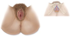
Labia Minora
The labia minora are thin folds of skin situated medial to the labia majora. They form the prepuce (hood) over the clitoris anteriorly and the frenulum posteriorly. These structures help protect the vaginal and urethral openings.

Vestibule
The vestibule is the space between the labia minora. It contains the urethral meatus, vaginal orifice, and hymen.

Urethral Meatus
The urethral meatus is the external opening of the urethra, located in the anterior portion of the vestibule, just posterior to the clitoris.

Vaginal Orifice
The vaginal orifice is the external opening to the vagina, situated posterior to the urethral meatus.
Hymen
The hymen is a thin fold of mucous membrane that partially encircles the vaginal orifice. It may partially occlude the vaginal opening until ruptured.

Clitoris
The clitoris is located at the anterior portion of the vestibule and is partially covered by the prepuce. The glans is the exposed distal end. The clitoris is highly innervated and plays a key role in female sexual arousal.

Lesser Vestibular Glands (Skene’s Glands)
These are an anterior pair of glands located on either side of the urethral meatus. They secrete mucus for lubrication.
Greater Vestibular Glands (Bartholin’s Glands)
Located posteriorly on either side of the vaginal orifice and covered by the bulbospongiosus muscle, these glands secrete mucus to lubricate the vestibule during sexual arousal.

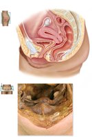
Internal Female Genitalia
Vagina
The vagina is a muscular tube extending from the perineal region superiorly to the uterus, situated between the urinary bladder and the rectum. It serves as the passageway for menstrual flow, intercourse, and childbirth.
Rugae
The rugae are folds of the mucous membrane lining the interior of the vagina, allowing for expansion during intercourse and childbirth.

Uterine Cervix
The uterine cervix projects inferiorly and posteriorly into the vagina, forming the lower part of the uterus.

Fornix
The fornix is the recess formed by the projection of the uterine cervix into the vagina.

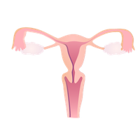
Uterus
The uterus is a pear-shaped, hollow organ located superior to the vagina and urinary bladder. It is divided into three main parts: the fundus, body, and cervix.

Fundus: The dome-shaped region superior to the uterine cavity.
Body: The main portion, surrounding the uterine cavity and continuous with the cervix inferiorly.
Cervix: The narrowed, inferior region of the uterus.

Cervical Canal and External Os
The cervical canal is a narrowed continuation of the uterine cavity that runs through the cervix. The external os is the opening of the cervical canal into the vagina.


Layers of the Uterus
Endometrium: Highly vascular inner lining that is shed during menstruation.
Myometrium: Thick middle layer of smooth muscle responsible for uterine contractions.
Perimetrium: The outermost layer, part of the peritoneum, which helps form the broad ligament.


Uterine Tubes (Fallopian Tubes)
The uterine tubes connect the uterus to the peritoneal cavity near each ovary. They are essential for the transport of ova from the ovaries to the uterus and are the typical site of fertilization.
Infundibulum: The expanded lateral end of the tube.
Ostium: The opening of the infundibulum next to the ovary.
Fimbriae: Finger-like projections that surround the ostium and help capture the ovulated oocyte.

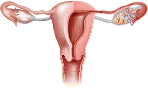

Ovaries
The ovaries are almond-shaped glands attached to the broad ligament. They produce oocytes (eggs) and secrete hormones such as estrogen and progesterone.
Tunica Albuginea: A thin fibrous capsule surrounding the ovary.
Stroma: The tissue within the tunica albuginea, divided into the medulla (highly vascular center) and cortex (outer region containing follicles).
Follicles: Round epithelial vesicles containing primary oocytes capable of developing into eggs.
Ligaments of the Female Reproductive System
Overview
Several ligaments support the female reproductive organs, maintaining their position within the pelvic cavity.
Broad Ligament: A double layer of peritoneum that hangs from the uterine tubes and supports the uterus, ovaries, and uterine tubes. It is divided into the mesometrium, mesovarium, and mesosalpinx.
Suspensory Ligament: The thickened superior edge of the broad ligament that attaches the ovary to the lateral pelvic wall.
Ovarian Ligament: Suspends the ovary from the uterus.
Round Ligament: Extends from the uterus, passes through the inguinal canal, and attaches to the connective tissue of the labia majora.
