 Back
BackANP College Study Guide: The Heart (Structure, Function, and Physiology)
Study Guide - Smart Notes
 Tailored notes based on your materials, expanded with key definitions, examples, and context.
Q1. What are the superior and inferior chambers of the heart called?
Background
Topic: Heart Anatomy
This question tests your knowledge of the basic structural divisions of the heart, specifically the names and locations of its chambers.
Key Terms:
Superior chambers: Located at the top of the heart.
Inferior chambers: Located at the bottom of the heart.
Step-by-Step Guidance
Recall that the heart has four chambers: two on the top and two on the bottom.
The chambers on the top are responsible for receiving blood.
The chambers on the bottom are responsible for pumping blood out of the heart.
Think about the anatomical terms used for these chambers.
Try solving on your own before revealing the answer!
Final Answer: Superior chambers are atria; inferior chambers are ventricles.
The atria are the upper chambers that receive blood, while the ventricles are the lower chambers that pump blood out of the heart.
Q2. What is the name of the wall that separates the right and left sides of the heart?
Background
Topic: Heart Anatomy
This question is about the internal structure of the heart, specifically the wall that divides the heart into right and left sides.
Key Terms:
Septum: A wall or partition separating two chambers.
Step-by-Step Guidance
Recall the term used for a wall that divides two areas in anatomy.
Think about the specific name for the wall in the heart that separates the right and left sides.
Consider if there are different types of septa in the heart (e.g., atrial, ventricular).
Try solving on your own before revealing the answer!
Final Answer: Septum
The septum is the wall that separates the right and left sides of the heart. There are both atrial and ventricular septa.
Q3. What is the function of the heart valves?
Background
Topic: Heart Physiology
This question tests your understanding of how the heart maintains unidirectional blood flow and prevents backflow.
Key Terms:
Valve: A structure that opens and closes to control the direction of blood flow.
Backflow: The reverse flow of blood, which valves prevent.
Step-by-Step Guidance
Recall the main purpose of valves in any circulatory system.
Think about what happens when the heart contracts and relaxes.
Consider how valves help maintain efficient circulation.
Try solving on your own before revealing the answer!
Final Answer: Heart valves prevent backflow and ensure blood flows in one direction.
Valves open and close in response to pressure changes, maintaining unidirectional flow through the heart.
Q4. What are the two main circuits of blood flow in the body?
Background
Topic: Circulatory System
This question is about the two major pathways that blood takes as it circulates through the body and heart.
Key Terms:
Pulmonary circuit: Carries blood between the heart and lungs.
Systemic circuit: Carries blood between the heart and the rest of the body.
Step-by-Step Guidance
Recall the two main destinations for blood leaving the heart.
Think about which circuit is responsible for oxygenating blood.
Consider which circuit delivers oxygenated blood to tissues.
Try solving on your own before revealing the answer!
Final Answer: Pulmonary and systemic circuits
The pulmonary circuit sends blood to the lungs for oxygenation, while the systemic circuit delivers oxygenated blood to the body.
Q5. Identify the structures labeled A–H in the heart conduction system diagram.
Background
Topic: Cardiac Conduction System
This question tests your ability to identify the anatomical components of the heart's electrical conduction system, which coordinates the heartbeat.
Key Terms:
SA node: Sinoatrial node, the heart's pacemaker.
AV node: Atrioventricular node, delays the impulse.
Bundle branches: Pathways for electrical signals.
Purkinje fibers: Distribute the impulse to ventricles.
Step-by-Step Guidance
Examine the diagram and locate the SA node, which is usually at the top of the right atrium.
Identify the AV node, found near the lower part of the right atrium.
Trace the pathway from the AV node to the bundle of His and then to the bundle branches.
Locate the Purkinje fibers, which spread throughout the ventricles.
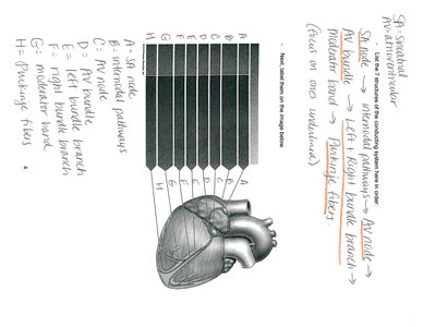
Try solving on your own before revealing the answer!
Final Answer: A. SA node, B. Internodal pathways, C. AV node, D. Bundle of His, E. Right bundle branch, F. Left bundle branch, G. Purkinje fibers, H. Moderator band
These structures are key components of the heart's electrical system, ensuring coordinated contraction.
Q6. What happens during systole and diastole in the cardiac cycle?
Background
Topic: Cardiac Cycle
This question is about the phases of the heartbeat, specifically what occurs during contraction (systole) and relaxation (diastole).
Key Terms:
Systole: Period of ventricular contraction.
Diastole: Period of ventricular relaxation.
Step-by-Step Guidance
Recall what happens to the ventricles during systole.
Think about the movement of blood during this phase.
Consider what happens during diastole and how the heart refills with blood.
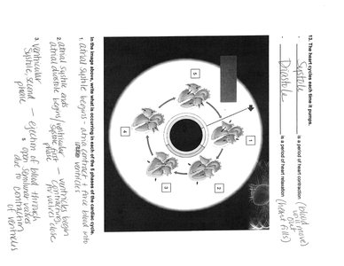
Try solving on your own before revealing the answer!
Final Answer: Systole is ventricular contraction (blood ejected); diastole is ventricular relaxation (heart refills).
During systole, the ventricles contract and push blood out; during diastole, they relax and fill with blood.
Q7. What is the role of the medulla oblongata in heart function?
Background
Topic: Neural Regulation of the Heart
This question tests your understanding of how the nervous system controls heart rate and function.
Key Terms:
Medulla oblongata: Part of the brainstem that regulates autonomic functions.
Autonomic nervous system: Controls involuntary actions like heart rate.
Step-by-Step Guidance
Recall the location and function of the medulla oblongata.
Think about how the medulla oblongata influences heart rate and blood pressure.
Consider the pathways through which the medulla communicates with the heart.
Try solving on your own before revealing the answer!
Final Answer: The medulla oblongata regulates heart rate and blood pressure via autonomic control.
It sends signals through the sympathetic and parasympathetic nervous systems to adjust cardiac function.
