 Back
BackAPK2105C Exam 1 UGA TA Review Material
Study Guide - Smart Notes
 Tailored notes based on your materials, expanded with key definitions, examples, and context.
Chapter 1: Introduction to Physiology
Definitions and Scope
Physiology is the study of body functions, while anatomy focuses on body structures. Applied physiology examines how structure and function are interrelated. Understanding physiology requires knowledge of the hierarchical organization of the body, from molecules to the whole organism.
Anatomy: Study of body structures.
Physiology: Study of body functions.
Applied Physiology: Relationship between structure and function.
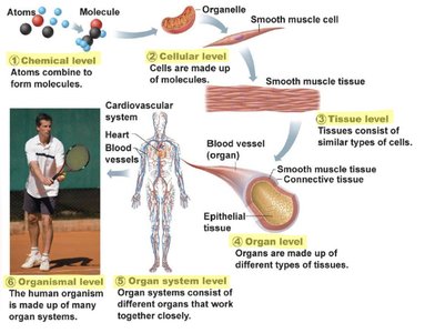
Hierarchy of Structural Organization
The human body is organized into several levels, each building upon the previous:
Chemical Level: Atoms combine to form molecules.
Cellular Level: Cells are made up of molecules and are the smallest living units.
Tissue Level: Tissues consist of similar types of cells working together.
Organ Level: Organs are made up of different types of tissues.
Organ System Level: Organ systems consist of different organs that work closely together.
Organismal Level: The human organism is made up of many organ systems.
Cells and Tissues
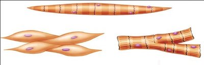
Cells are the fundamental units of life. Tissues are groups of cells with similar structure and function. The four primary tissue types are:
Nervous Tissue: Contains neurons (for communication and control) and neuroglia (support cells).
Muscle Tissue: Specialized for movement; also involved in thermoregulation, extensibility, elasticity, contractility, and excitability.
Epithelial Tissue: Sheet-like arrangement of cells; forms glands and lines surfaces.
Connective Tissue: Most abundant and diverse; provides support and structure.
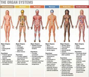
Organs and Organ Systems
Organs are composed of multiple tissue types working together for specific functions. Organ systems are groups of organs that perform related functions. Many organ systems share organs and overlap in function.
Epithelium and Semipermeable Membranes
One layer of epithelial tissue separates the internal environment from the external environment. Semipermeable membranes regulate the movement of substances between fluid compartments in the body.
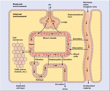
Homeostasis
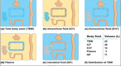
Homeostasis is the maintenance of constant internal conditions (especially in the extracellular fluid, ECF). All organ systems except the reproductive system contribute to homeostasis. The body regulates ECF to ensure optimal conditions for cellular function.
Key regulated variables: Composition, temperature, and volume of ECF.
Feedback Mechanisms
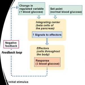
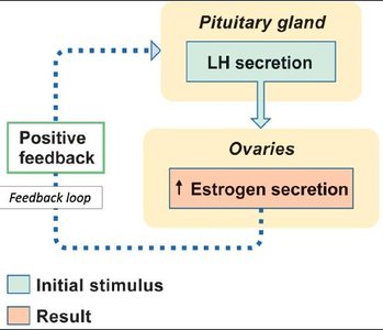
Feedback mechanisms maintain homeostasis:
Negative Feedback: Reverses a change to maintain stability (e.g., blood glucose regulation).
Positive Feedback: Amplifies a change to drive a process to completion (e.g., LH surge in ovulation).
Chapter 2: The Cell Structure and Function
Biological Macromolecules
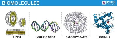
There are four main types of large biological macromolecules essential for life:
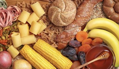
Carbohydrates: Polymers of monosaccharides; main energy source.
Lipids: Nonpolar, hydrophobic molecules; include fats, oils, and steroids.
Proteins: Polymers of amino acids; perform structural, enzymatic, and regulatory functions.
Nucleic Acids: Polymers of nucleotides; store and transfer genetic information.
Carbohydrates
Carbohydrates are polar molecules that dissolve easily in water. They are classified as:
Monosaccharides: Simple sugars (e.g., glucose).
Disaccharides: Two monosaccharides covalently bonded (e.g., sucrose, lactose).
Polysaccharides: Long chains of monosaccharide subunits (e.g., starch, glycogen).
Lipids
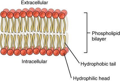
Lipids are nonpolar and insoluble in water. Major types include:
Triglycerides: Glycerol + 3 fatty acids; main form of stored energy.
Phospholipids: Amphipathic molecules forming cell membranes.
Steroids: Cholesterol-based molecules; precursors for hormones.
Eicosanoids: Signaling molecules derived from fatty acids.
Proteins
Proteins are polymers of amino acids joined by peptide bonds. They have four levels of structure:
Primary: Sequence of amino acids.
Secondary: Alpha-helices and beta-sheets formed by hydrogen bonding.
Tertiary: 3D folding due to R group interactions.
Quaternary: Multiple polypeptide chains interacting.
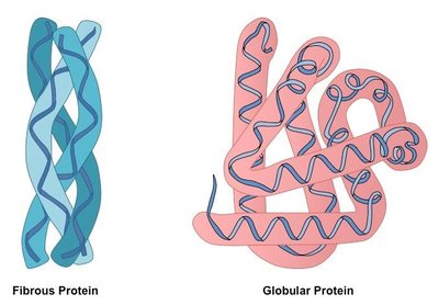
Fibrous Proteins: Structural roles (e.g., collagen).
Globular Proteins: Functional roles (e.g., enzymes, hormones).
Nucleic Acids and Nucleotides
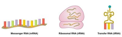
Nucleotides are composed of a nitrogenous base, a five-carbon sugar, and a phosphate group. DNA and RNA are nucleic acids responsible for genetic information storage and transfer.
DNA: Double helix, stores genetic code.
RNA: Single-stranded, involved in protein synthesis.
Pyrimidines: Cytosine, thymine (uracil in RNA).
Purines: Adenine, guanine.
Membranous Organelles
Key organelles include:
Rough Endoplasmic Reticulum (ER): Protein synthesis (with ribosomes).
Smooth ER: Lipid and steroid synthesis, calcium storage.
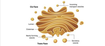
Golgi Apparatus: Protein modification, packaging, and shipping.
Mitochondria: ATP production via aerobic respiration.
Lysosomes: Digestive enzymes for autophagy.
Peroxisomes: Breakdown of toxic substances, produce and degrade H2O2.
The Cytoskeleton
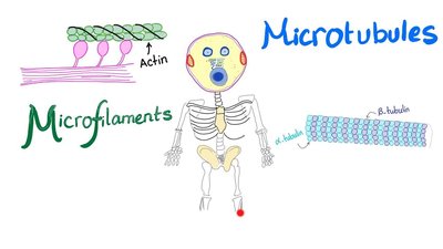
The cytoskeleton provides structural support and facilitates movement:
Microfilaments: Actin; smallest, involved in cell shape and movement.
Intermediate Filaments: Myosin, keratin; structural support.
Microtubules: Tubulin; largest, form cilia and flagella.
Cell-to-Cell Adhesions
Cells are connected by specialized junctions:
Tight Junctions: Seal cells to prevent leakage (e.g., in the bladder).
Desmosomes: Anchor cells together, resist stretching (e.g., skin, heart).
Gap Junctions: Allow communication via connexons (e.g., cardiac muscle).
Chapter 3: Cell Metabolism
Metabolism and Metabolic Pathways
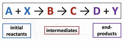
Metabolism is the sum of all chemical reactions in the cell. It includes:
Anabolism: Synthesis of large molecules from smaller ones (requires energy).
Catabolism: Breakdown of large molecules into smaller ones (releases energy).
Types of Metabolic Reactions
Hydrolysis: Splitting molecules with water (catabolic).
Condensation: Joining molecules, producing water (anabolic).
Phosphorylation: Addition of phosphate group.
Dephosphorylation: Removal of phosphate group.
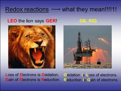
Oxidation: Loss of electrons.
Reduction: Gain of electrons.
Energy Transfer in Cells
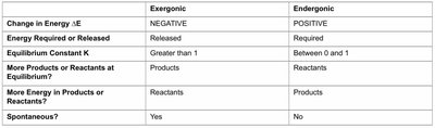
Energy is the capacity to do work. In cells, energy is transferred through metabolic reactions. Energy can be kinetic (motion) or potential (stored in chemical bonds). The direction and spontaneity of reactions are determined by changes in free energy ().
Exergonic Reactions: Release energy (), spontaneous.
Endergonic Reactions: Require energy (), non-spontaneous.
Enzymes and Reaction Rates
Enzymes are biological catalysts that speed up reactions by lowering activation energy. They are not consumed in the reaction and are highly specific for their substrates.
Factors Affecting Enzyme Activity: Temperature, pH, cofactors, coenzymes, substrate concentration, enzyme concentration, and affinity for substrate.
Induced Fit Model
The induced fit model describes how the enzyme's active site changes shape to fit the substrate, allowing for catalysis and product formation.
Cofactors and Coenzymes
Cofactors are non-protein helpers required for enzyme activity. Coenzymes are organic cofactors, often derived from vitamins, that carry chemical groups between reactions.
Examples: NADH (electron carrier), CoA (acyl carrier), FAD (electron carrier).
Enzyme Regulation
Allosteric Regulation: Modulator binds to a site other than the active site, altering enzyme activity.
Covalent Regulation: Covalent modification (e.g., phosphorylation) activates or deactivates the enzyme.
Feedback Inhibition
In feedback inhibition, the end product of a metabolic pathway inhibits an earlier enzyme, preventing overproduction. The inhibited enzyme is often called the "rate-limiting enzyme."
ATP and Energy Metabolism
ATP is the primary energy currency of the cell. It is produced by substrate-level phosphorylation (direct transfer of phosphate) and oxidative phosphorylation (electron transport chain, requires oxygen).
Glucose Oxidation
Glucose is oxidized in several stages to produce ATP:
Glycolysis: Occurs in the cytoplasm; anaerobic; produces pyruvate, ATP, and NADH.
Linking Step: Converts pyruvate to acetyl-CoA.
Krebs Cycle: Occurs in the mitochondrial matrix; produces NADH, FADH2, ATP, and CO2.
Oxidative Phosphorylation: Electron transport chain and chemiosmosis; produces most ATP.
Alternative Fuels and Storage
Fats: Broken down via lipolysis for energy.
Proteins: Broken down via proteolysis when necessary.
Glycogen: Storage form of glucose in liver and muscle.
The liver plays a key role in maintaining blood glucose by storing and releasing glucose as needed.
