 Back
BackAutonomic Nervous System: Parasympathetic Division, Neurotransmitters, Dual Innervation, and Higher-Order Functions
Study Guide - Smart Notes
 Tailored notes based on your materials, expanded with key definitions, examples, and context.
Parasympathetic Division of the Autonomic Nervous System
Major Effects of Parasympathetic Activation
The parasympathetic division is responsible for conserving energy and promoting 'rest-and-digest' functions. Its activation leads to several physiological changes that support maintenance and restoration of the body.
Constriction of pupils and focusing of the lenses for near vision
Stimulation of digestive gland secretion (salivary, gastric, intestinal, duodenal, pancreatic, and hepatic glands)
Hormone secretion that promotes nutrient absorption and utilization
Changes in blood flow and glandular activity associated with sexual arousal
Increased smooth muscle activity along the digestive tract
Stimulation and coordination of defecation
Contraction of the urinary bladder during urination
Constriction of respiratory passageways
Reduction in heart rate and force of contraction
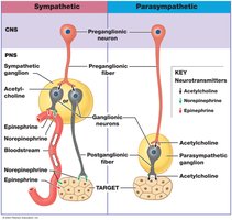
Parasympathetic Neurotransmitters and Receptors
Neurotransmitter Release and Inactivation
All parasympathetic neurons release acetylcholine (ACh) as their neurotransmitter. The effects of ACh depend on the type of receptor present on the postsynaptic cell and the second messenger systems involved.
Most ACh is rapidly inactivated at the synapse by acetylcholinesterase (AChE).
ACh that diffuses into surrounding tissues is inactivated by tissue cholinesterase.
Cholinergic Receptors
Cholinergic receptors are proteins that bind ACh and mediate its effects. There are two main types:
Nicotinic receptors: Found on postganglionic neurons of both sympathetic and parasympathetic divisions, and at neuromuscular junctions in the somatic nervous system. These are chemically gated Na+ channels; activation by ACh causes excitation of the postganglionic neuron.
Muscarinic receptors: Located at target organs and tissues of the parasympathetic division and at some cholinergic synapses in the sympathetic division. These are G protein-coupled receptors, and their response can be excitatory or inhibitory depending on the enzymes activated or inactivated.
Summary Comparison of Sympathetic and Parasympathetic Divisions
Structural and Functional Differences
Sympathetic Division:
Widespread effects due to extensive divergence
Short preganglionic and long postganglionic fibers
Preganglionic neurons release ACh; most postganglionic fibers release norepinephrine (NE), some release ACh or nitric oxide (NO)
Receptors: Nicotinic cholinergic on postganglionic neurons, adrenergic and some muscarinic cholinergic on target cells
Parasympathetic Division:
More specific, localized effects
Ganglionic neurons located in or near target organs
All neurons are cholinergic
Receptors: Nicotinic cholinergic on postganglionic neurons, muscarinic cholinergic on target cells
Effects are brief and restricted to specific sites
Dual Innervation and Autonomic Tone
Dual Innervation
Most vital organs receive input from both sympathetic and parasympathetic divisions, often with opposing effects. This arrangement allows for precise regulation of organ function.
Parasympathetic postganglionic fibers travel via cranial nerves to peripheral destinations.
Sympathetic innervation reaches the same structures from the superior cervical ganglia.
Autonomic plexuses are nerve networks formed by mingled sympathetic and parasympathetic fibers, traveling with blood and lymphatic vessels to visceral organs.
Autonomic Tone
Autonomic motor neurons maintain a resting level of activity, known as autonomic tone. This allows for a greater range of control by increasing or decreasing activity as needed.
Significant where dual innervation occurs, but especially important where only one division innervates an organ (e.g., blood vessels are only under sympathetic control).
Sympathetic tone keeps blood vessels partially contracted; decreased NE release causes vasodilation.
Example: Heart Regulation
ACh from parasympathetic fibers slows heart rate.
NE from sympathetic fibers accelerates heart rate.
Both are released continuously, producing autonomic tone; parasympathetic division dominates at rest.
Regulation of Autonomic Functions
Visceral Reflexes
Visceral reflexes are polysynaptic reflexes that provide automatic motor responses in glands and nonskeletal muscle organs. They can be modified by higher centers, especially the hypothalamus.
Components of a visceral reflex arc:
Receptor
Sensory neuron
Processing center (one or more interneurons)
Two visceral motor neurons (preganglionic and postganglionic)
Peripheral effector
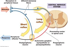
Types of Visceral Reflexes
Long reflexes: Coordinate activities of entire organs; sensory input is processed in the CNS, and motor commands are sent via the ANS.
Short reflexes: Bypass the CNS, controlling activity in one part of an organ; important for localized digestive tract functions.
Enteric Nervous System
The enteric nervous system is capable of controlling digestive functions independently of the CNS. It consists of ganglia in the walls of the digestive tract containing sensory neurons, interneurons, and motor neurons, forming extensive nerve nets.
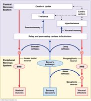
Higher Levels of Autonomic Control
Medulla oblongata coordinates complex reflexes (salivation, swallowing, digestive secretions, movement, urinary function).
Regulated by the hypothalamus.
Integration of ANS and SNS Activities
The autonomic (ANS) and somatic (SNS) nervous systems have parallel organization and are integrated at the brainstem level. Both are under the control of higher brain centers.
Higher-Order Functions
Characteristics of Higher-Order Functions
Require the cerebral cortex
Involve both conscious and unconscious information processing
Subject to adjustment over time (not innate)
Memory Types and Storage
Fact memories: Specific bits of factual information
Skill memories: Learned motor behaviors, often incorporated unconsciously with repetition
Short-term memories: Briefly retained, immediately accessible
Long-term memories: More permanent; divided into secondary (fade with time) and tertiary (lifelong) memories
Memory consolidation is the process of converting short-term memories into long-term memories, often requiring repetition.
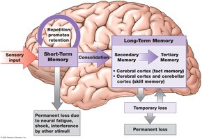
Brain Regions Involved in Memory
Amygdaloid body and hippocampus: Essential for memory consolidation; damage impairs new long-term memory formation
Cerebral cortex: Stores most long-term memories; association areas handle specific types (visual, auditory, etc.)
Some memories depend on single neurons ('grandmother cells')
Memory Engrams and Factors Affecting Memory Formation
Memory engram: A neural circuit corresponding to a single long-term memory, formed through experience and repetition
Factors: Nature, intensity, frequency of stimulus; CNS stimulants (caffeine, nicotine); hippocampal activity; NMDA receptor involvement
States of Consciousness and Sleep
Conscious state: Awareness and attention to external events
Unconscious state: Unresponsive, can be light or deep; degree of wakefulness reflects CNS activity
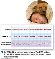
Sleep Types
Deep sleep (NREM): Body relaxes, cerebral cortex activity minimal, vital signs decrease
REM sleep: Active dreaming, rapid eye movements, fluctuating vital signs, muscle inhibition, EEG resembles awake state
Regulation of Sleep–Wake Cycles
Sleep ends with activation of the reticular formation and reticular activating system (RAS)
Awake state maintained by positive feedback from cerebral cortex and basal nuclei
Regulation involves 'dueling' nuclei in the brainstem: one group stimulates RAS with norepinephrine (NE) for alertness, another depresses RAS with serotonin for deep sleep
