 Back
Axial Musculature: Head, Neck, and Trunk Study Notes
Study Guide - Smart Notes
 Tailored notes based on your materials, expanded with key definitions, examples, and context.
Axial Musculature
Introduction
The axial musculature includes the muscles of the head, neck, and trunk. These muscles provide strength, stability, and facilitate specialized functions such as facial expression, mastication (chewing), breathing, and posture. Understanding their attachments, actions, and innervation is essential for comprehending their roles in human movement and physiology.
Muscles of the Head
Facial Expression Muscles
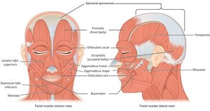
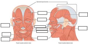
Muscles of facial expression originate on the skull and insert into the skin, allowing for a wide range of facial movements. Their unique insertions enable the skin to move more than in other body regions, creating expressions.
Orbicularis oris: Circular muscle that closes and protrudes the lips.
Orbicularis oculi: Circular muscle that closes the eyelids.
Occipitofrontalis: Raises the scalp and eyebrows; consists of frontal and occipital bellies connected by the epicranial aponeurosis.
Zygomaticus major and minor: Elevate the corners of the mouth (smiling/laughing).
Risorius: Draws the angle of the mouth laterally (grinning).
Levator labii superioris: Raises and protrudes the upper lip.
Mentalis: Protrudes the lower lip and elevates the chin (pouting).
Depressor labii inferioris: Depresses the lower lip.
Buccinator: Compresses the cheek, assists in chewing, whistling, and blowing.
Muscles of Mastication
These muscles are responsible for moving the mandible during chewing and speaking.
Masseter: Elevates the mandible to close the mouth; primary muscle for chewing.
Temporalis: Elevates and retracts the mandible.
Buccinator: Assists in holding food between teeth during chewing.
Table: Muscles of the Head
Muscle Name | Primary Action | Origin | Insertion | Innervation |
|---|---|---|---|---|
Frontalis | Raises eyebrows, draws scalp anteriorly | Galea aponeurotica | Subcutaneous tissue of eyebrows | Facial nerve (VII) |
Temporalis | Elevates (closes) mandible | Temporal fossa | Coronoid process and ramus of mandible | Trigeminal nerve (V) |
Masseter | Elevates (closes) mandible | Zygomatic arch | Angle and ramus of mandible | Trigeminal nerve (V) |
Orbicularis oculi | Closes eyelid | Frontal and maxilla on medial margin of orbit | Skin of eyelid | Facial nerve (VII) |
Orbicularis oris | Closes lips | Modiolus of mouth | Skin near lips | Facial nerve (VII) |
Risorius | Abducts corner of mouth | Fascia of masseter, zygomatic bone | Modiolus at angle of mouth | Facial nerve (VII) |
Mentalis | Protrudes lower lip | Anterior mandible | Skin of chin below lower lip | Facial nerve (VII) |
Buccinator | Compresses cheek | Maxilla and mandible near molar teeth | Orbicularis oris | Facial nerve (VII) |
Zygomaticus (major & minor) | Elevates corner of mouth | Zygomatic bone | Superolateral angle of mouth, upper lip muscles | Facial nerve (VII) |
Depressor labii inferioris | Depresses inferior lip | Mandible | Muscles and skin of inferior lip | Facial nerve (VII) |
Levator labii superioris | Elevates superior lip, flares nostril | Maxilla and zygomatic bones | Muscles of superior lip | Facial nerve (VII) |
Muscles of the Neck and Back
Anterior Neck Muscles
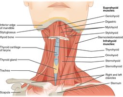
These muscles assist in swallowing and speech by controlling the larynx and hyoid bone. They are categorized by their position relative to the hyoid bone.
Digastric: Elevates the hyoid bone and depresses the mandible.
Mylohyoid: Forms the floor of the oral cavity, elevates the hyoid, and depresses the tongue.
Omohyoid: Depresses the hyoid bone.
Lateral and Posterior Neck Muscles
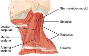
These muscles balance, move, and rotate the head. Unilateral contraction rotates the head, while bilateral contraction flexes or extends it.
Sternocleidomastoid: Abducts, rotates, and flexes the head; divides the neck into anterior and posterior triangles.
Platysma: Depresses the mandible and lower lip, tenses skin of the neck.
Scalene muscles (anterior, middle, posterior): Flex and rotate the neck, elevate ribs during deep inhalation.
Levator scapulae: Elevates the scapula and assists in neck movement.
Muscles of the Back
The back muscles stabilize and move the vertebral column. The erector spinae group is the primary extensor, controlling flexion, lateral flexion, and rotation of the vertebral column.
Iliocostalis: Lateral column of erector spinae.
Longissimus: Intermediate column.
Spinalis: Medial column.
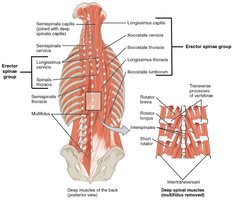
Table: Muscles of the Neck and Back
Muscle Name | Primary Action | Origin | Insertion | Innervation |
|---|---|---|---|---|
Levator scapulae | Elevates scapula, abducts and rotates neck | Vertebrae C1-4 | Upper vertebral border of scapula | Spinal nerves C3-5, posterior scapular nerve |
Scalenes | Flex and rotate neck, elevate ribs 1 and 2 | Transverse process of cervical vertebrae | Ribs 1 and 2 | Spinal nerves C3-8 |
Sternocleidomastoid | Abduct, rotate, and flex head | Sternum, clavicle | Mastoid process of temporal bone | Spinal nerves C2-4, accessory nerve (XI) |
Omohyoid | Depress hyoid bone | Superior border of scapula | Hyoid bone | Spinal nerves C1-3 |
Platysma | Depress inferior lip, open jaw | Fascia covering pectoralis major and deltoid | Mandible and skin of inferior face | Facial nerve (VII) |
Digastric | Open mouth; depress mandible | Inferior, distal margin of mandible, mastoid notch of temporal | Hyoid bone | Trigeminal (V) and facial (VII) nerves |
Mylohyoid | Elevate hyoid bone and tongue | Inferior margin of mandible | Hyoid bone | Trigeminal nerve (V) |
Erector spinae | Extend and rotate vertebral column and head | Nuchal ligament, thoracic/lumbar vertebrae, sacrum, ilium, ribs 3-12 | All ribs, thoracic vertebrae, temporal bones | Dorsal rami of spinal nerves |
Muscles That Position the Pectoral Girdle
The pectoral girdle consists of the clavicle, scapula, and proximal humerus, stabilized by muscles on the anterior and posterior thorax. These muscles allow for a wide range of arm movements.
Serratus anterior: Abducts scapula, moves it away from the spine.
Pectoralis minor: Depresses the scapula.
Trapezius: Extends and abducts the head, rotates and adducts scapula.
Rhomboid major and minor: Adduct the scapula.
Pectoralis major: Flexes, adducts, and medially rotates the humerus.
Latissimus dorsi: Extends, adducts, and medially rotates the humerus.
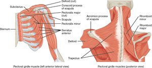
Table: Muscles of the Pectoral Girdle
Muscle Name | Primary Action | Origin | Insertion | Innervation |
|---|---|---|---|---|
Pectoralis major | Flex, adduct, medially rotate humerus | Clavicle, sternum, ribs 1-7 | Crest of greater tubercle of humerus | Medial/lateral pectoral nerves |
Pectoralis minor | Depress glenoid cavity | Ribs 3-5 | Coracoid process of scapula | Medial/lateral pectoral nerves |
Trapezius | Extend/abduct head, rotate/adduct scapula | Occipital bone, nuchal ligament, vertebrae C7-T4 | Clavicle, acromion, spine of scapula | Accessory nerve (XI) |
Latissimus dorsi | Extend, adduct, medially rotate humerus | Vertebrae T7-12, L1-5, ilium, ribs 10-12 | Intertubercular sulcus of humerus | Thoracodorsal nerve |
Rhomboid major | Adduct scapula | Spines of vertebrae T2-5 | Medial border of scapula | Dorsal scapular nerve |
Rhomboid minor | Adduct scapula | Nuchal ligament, spines of vertebrae C7-T1 | Medial border of scapula | Dorsal scapular nerve |
Muscles of the Thorax
Intercostal Muscles
These muscles facilitate breathing by altering the size of the thoracic cavity.
External intercostals: Elevate the ribs during inspiration.
Internal intercostals: Depress the ribs during forced expiration.
Innermost intercostals: Assist internal intercostals.
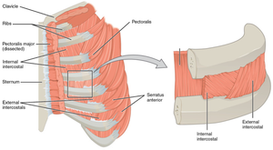
Diaphragm
The diaphragm is the primary muscle of respiration, separating the thoracic and abdominal cavities. Its contraction increases thoracic volume, causing inhalation; relaxation causes exhalation. It also plays a role in increasing intra-abdominal pressure during defecation, urination, and childbirth (Valsalva maneuver).
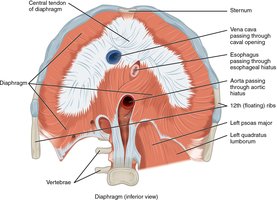
Table: Muscles of the Thorax
Muscle Name | Primary Action | Origin | Insertion | Innervation |
|---|---|---|---|---|
External intercostals | Elevate ribs (inspiration) | Inferior border of ribs 1-11 | Superior border of next inferior rib | Intercostal nerves |
Internal intercostals | Depress ribs (expiration) | Superior border of ribs 2-12 | Inferior border of next superior rib | Intercostal nerves |
Diaphragm | Inspiration | Xiphoid process, ribs 7-12, lumbar vertebrae | Central tendon of diaphragm | Phrenic nerve |
Muscles of the Abdomen
The abdominal muscles stabilize the trunk, maintain posture, and protect internal organs. They are arranged in layers with different fiber orientations for strength and flexibility.
Rectus abdominis: Flexes the vertebral column and compresses the abdomen.
External abdominal oblique: Compresses the abdominal wall, laterally rotates the waist.
Internal abdominal oblique: Compresses the abdominal wall, laterally rotates the waist.
Transverse abdominis: Compresses the abdominal wall.
Quadratus lumborum: Extends and abducts the vertebral column.
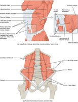
Table: Muscles of the Abdomen
Muscle Name | Primary Action | Origin | Insertion | Innervation |
|---|---|---|---|---|
External abdominal oblique | Compress abdominal wall, laterally rotate waist | Ribs 5-12 | Iliac crest, pubis | Intercostal nerves T7-12 |
Internal abdominal oblique | Compress abdominal wall, laterally rotate waist | Inguinal ligament, iliac crest | Pubis, ribs 10-12 | Intercostal nerves T7-12, spinal nerve L1 |
Transverse abdominal | Compress abdominal wall | Inguinal ligament, iliac crest, costal cartilages 7-12 | Linea alba, pubis | Intercostal nerves T7-12, spinal nerve L1 |
Rectus abdominis | Flex vertebral column, compress abdominal wall | Crest of pubis, pubic symphysis | Costal cartilages 5-7, xiphoid process | Intercostal nerves T6-12 |
Serratus anterior | Abduct scapula | Ribs 1-8 | Vertebral border and inferior angle of scapula | Long thoracic nerve |
Quadratus lumborum | Extend and abduct vertebral column | Iliac crest | Vertebrae L1-4, rib 12 | L1-4, T12 |
Summary Table: Key Muscle Actions and Innervations
Muscle | Action | Innervation |
|---|---|---|
Temporalis | Elevates and retracts mandible | Trigeminal nerve (V) |
Masseter | Elevates mandible | Trigeminal nerve (V) |
Zygomaticus (major & minor) | Elevates corners of mouth | Facial nerve (VII) |
Sternocleidomastoid | Abducts, rotates, flexes head | Accessory nerve (XI), spinal nerves C2-4 |
Rectus abdominis | Flexes vertebral column, compresses abdomen | Intercostal nerves T6-12 |
Additional info:
The Valsalva maneuver is a coordinated action of the diaphragm and abdominal muscles to increase intra-abdominal pressure, aiding in defecation, urination, and childbirth.
The linea alba is a fibrous structure running down the midline of the abdomen, where the aponeuroses of the abdominal muscles meet.
