 Back
BackBIO163 Endocrine, Eye, and Ear Study Guidance
Study Guide - Smart Notes
 Tailored notes based on your materials, expanded with key definitions, examples, and context.
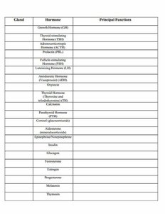
Q1. Complete the table for endocrine glands, hormones, principal functions, and hormone type (protein/steroid).
Background
Topic: Endocrine System
This question tests your knowledge of the major endocrine glands, the hormones they produce, the principal functions of those hormones, and whether each hormone is a protein or steroid based on its effect on target cells.
Key Terms:
Endocrine gland: An organ that secretes hormones directly into the bloodstream.
Hormone: A chemical messenger that regulates physiological processes.
Protein hormone: Usually acts via cell surface receptors.
Steroid hormone: Usually acts via intracellular receptors.
Step-by-Step Guidance
Review the list of glands and hormones in the table. Identify which gland produces each hormone.
For each hormone, research or recall its principal function (e.g., growth hormone stimulates growth and metabolism).
Determine whether each hormone is a protein or steroid. Protein hormones are typically peptides or glycoproteins, while steroid hormones are derived from cholesterol.
Add a column to the table to indicate the hormone type for each entry.
Fill in the missing hormones and their functions for any gaps in the table.
Try solving on your own before revealing the answer!
Final Answer:
Each row should include the gland, hormone, principal function, and whether the hormone is a protein or steroid. For example, Growth Hormone (GH) is produced by the anterior pituitary, stimulates growth, and is a protein hormone.
Continue this process for all hormones listed in the table.
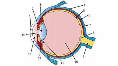
Q2. Identify and describe the parts of the eye in the diagram, then fill out the table with structure, physiology/function, and location.
Background
Topic: Special Senses – Eye Anatomy and Physiology
This question tests your ability to identify anatomical structures of the eye, describe their physiological functions, and specify their locations.
Key Terms:
Cornea: Transparent front part of the eye that refracts light.
Retina: Layer containing photoreceptors for vision.
Lens: Focuses light onto the retina.
Optic nerve: Transmits visual information to the brain.
Step-by-Step Guidance
Examine the diagram and identify each numbered structure (e.g., cornea, lens, retina).
For each structure, describe its main physiological function (e.g., the lens focuses light).
Specify the location of each structure within the eye (e.g., anterior, posterior, etc.).
Fill out the table with structure, function, and location for each numbered part.
Try solving on your own before revealing the answer!
Final Answer:
The completed table should list each structure (e.g., cornea, lens, retina), its function (e.g., refracts light, focuses light, detects light), and its location (e.g., anterior, posterior).
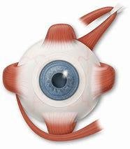
Q3. Label the external eye muscles, their function, and innervation in the diagram.
Background
Topic: Eye Muscles and Innervation
This question tests your knowledge of the extraocular muscles, their actions, and the cranial nerves that innervate them.
Key Terms:
Extraocular muscles: Muscles that move the eyeball.
Innervation: The cranial nerves that supply each muscle (III, IV, VI).
Step-by-Step Guidance
Identify each external eye muscle in the diagram (e.g., superior rectus, lateral rectus).
List the primary function of each muscle (e.g., elevation, abduction).
Determine which cranial nerve innervates each muscle (e.g., oculomotor, trochlear, abducens).
Label the diagram and fill out the table with muscle, function, and innervation.
Try solving on your own before revealing the answer!
Final Answer:
Each muscle should be labeled with its name, function, and innervating cranial nerve (e.g., lateral rectus – abduction – CN VI).
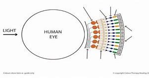
Q4. Label the photoreceptors in the diagram and list the function of each.
Background
Topic: Photoreceptors in the Retina
This question tests your understanding of the types of photoreceptors (rods and cones) and their functions in vision.
Key Terms:
Rod: Photoreceptor for low-light vision.
Cone: Photoreceptor for color and high-acuity vision.
Step-by-Step Guidance
Identify rods and cones in the diagram.
List the function of rods (night vision, peripheral vision).
List the function of cones (color vision, sharp vision).
Label the diagram and fill out the table with photoreceptor type and function.
Try solving on your own before revealing the answer!
Final Answer:
Rods are responsible for dim light vision; cones are responsible for color and detail.
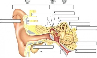
Q5. Label all structures in the ear diagram and list the function of each. Know the ossicles in order, organs for static/dynamic equilibrium, hearing sensation, and VIIIth cranial nerve divisions and function.
Background
Topic: Ear Anatomy and Physiology
This question tests your knowledge of the anatomical structures of the ear, their functions, the order of ossicles, and the role of the vestibulocochlear nerve (VIII).
Key Terms:
Ossicles: Malleus, incus, stapes (in order).
Static equilibrium: Controlled by the vestibule.
Dynamic equilibrium: Controlled by the semicircular canals.
Hearing: Controlled by the cochlea.
VIIIth cranial nerve: Vestibulocochlear nerve (hearing and balance).
Step-by-Step Guidance
Identify and label each structure in the ear diagram (outer, middle, inner ear).
List the function of each structure (e.g., cochlea – hearing, semicircular canals – dynamic equilibrium).
Recall the order of the ossicles (malleus, incus, stapes).
Identify the organs responsible for static and dynamic equilibrium.
Describe the divisions and function of the VIIIth cranial nerve.
Try solving on your own before revealing the answer!
Final Answer:
The labeled diagram should include all structures, their functions, the correct order of ossicles, and the role of the vestibulocochlear nerve.
