Back
BackBlood and the Cardiovascular System: Structure, Function, and Disorders
Study Guide - Smart Notes
 Tailored notes based on your materials, expanded with key definitions, examples, and context.
Blood and the Cardiovascular System
Plasma
Plasma is the straw-colored, sticky fluid portion of blood, making up about 55% of total blood volume. It is composed mostly of water and contains over 100 dissolved solutes, including gases, hormones, and electrolytes. Electrolytes are the most abundant solutes in plasma. Plasma proteins, primarily produced by the liver, are not used as fuel by cells but serve important functions such as maintaining osmotic pressure and transporting substances.
Albumin: The most abundant plasma protein (60%), produced by the liver. It acts as a carrier for molecules, a blood buffer, and is the major contributor to plasma osmotic pressure, helping to keep water in the bloodstream.
Formed Elements of Blood
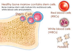
The formed elements of blood include erythrocytes (red blood cells), leukocytes (white blood cells), and platelets. Only leukocytes are complete cells; erythrocytes lack nuclei and organelles, and platelets are cell fragments. Most formed elements survive only a few days in the bloodstream and are replaced by stem cells in the bone marrow.
Erythrocytes: Specialized for transporting respiratory gases (oxygen and carbon dioxide).
Leukocytes: Crucial for defense against disease.
Platelets: Involved in blood clotting.
Erythrocytes (Red Blood Cells)
Structure and Function
Mature erythrocytes are biconcave discs lacking nuclei and organelles, essentially bags of hemoglobin and some antioxidant enzymes. Their shape provides a 30% greater surface area than spherical cells, facilitating efficient gas exchange. They generate ATP anaerobically, so they do not consume the oxygen they transport.
Over 97% of erythrocyte content (excluding water) is hemoglobin.
Each erythrocyte contains about 250 million hemoglobin molecules, allowing each cell to carry about 1 billion oxygen molecules.
Hemoglobin
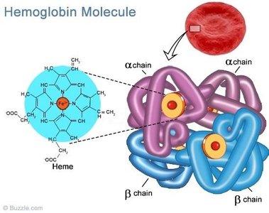
Hemoglobin is a protein that binds oxygen easily and reversibly. It consists of four polypeptide chains (globin), each bound to a heme group. Each heme contains an iron atom that can bind one oxygen molecule, so one hemoglobin molecule can transport four oxygen molecules.
Oxygen-bound hemoglobin is called oxyhemoglobin (ruby red).
When oxygen detaches, it becomes deoxyhemoglobin (dark red).
Erythropoiesis (Red Blood Cell Production)
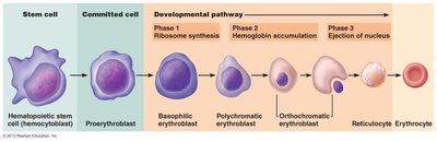
Erythropoiesis is the process of red blood cell formation, occurring in red bone marrow (axial skeleton, girdles, humerus, and femur). The process from hematopoietic stem cell to reticulocyte takes about 15 days, with an additional 2 days in the bloodstream to become fully mature.
Regulation of Erythropoiesis
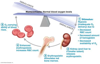
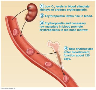
Erythropoiesis is regulated hormonally, primarily by erythropoietin (EPO), a glycoprotein hormone produced by the kidneys (and to a lesser extent, the liver). EPO stimulates the maturation of red blood cells in response to hypoxia (low oxygen levels).
Fate and Destruction of Erythrocytes
Erythrocytes have a lifespan of 100–120 days. They cannot synthesize new proteins, grow, or divide. As they age, they become fragile and are removed from circulation, primarily by macrophages in the spleen. Iron from hemoglobin is recycled, the heme group is converted to bilirubin, and globin is broken down into amino acids.
Erythrocyte Disorders
Anemia: Reduced oxygen-carrying capacity of blood, leading to fatigue, pallor, shortness of breath, and chills. Causes include blood loss, insufficient RBC production, or excessive RBC destruction.
Iron-deficiency anemia: Characterized by microcytes (small, pale RBCs) due to lack of iron.
Pernicious anemia: Autoimmune disorder affecting the elderly, due to vitamin B12 deficiency.
Renal anemia: Caused by lack of EPO, often due to kidney damage.
Aplastic anemia: Failure of red bone marrow, often due to drugs or viruses.
Hemolytic anemia: RBCs rupture or lyse prematurely.
Sickle-cell anemia: Abnormal hemoglobin causes RBCs to become spiky and rupture easily under low-oxygen conditions.
Leukocytes (White Blood Cells)
General Characteristics
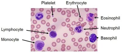
Leukocytes are less numerous than erythrocytes but are essential for defense against disease. They are complete cells with nuclei and organelles and can leave the bloodstream to enter tissues (diapedesis). Leukocytes are classified into two main categories: granulocytes and agranulocytes.
Granulocytes
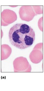
Neutrophils: Most abundant WBCs (50–70%), multilobed nucleus, very phagocytic, especially against bacteria.
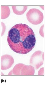
Eosinophils: Bilobed nucleus, red-staining granules, combat parasitic worms, involved in allergies and asthma.
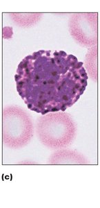
Basophils: Rarest WBCs, large granules containing histamine (inflammatory mediator).
Agranulocytes
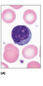
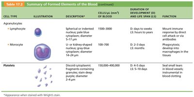
Lymphocytes: Large, dark nucleus, crucial for immunity. T cells attack infected/tumor cells; B cells produce antibodies.
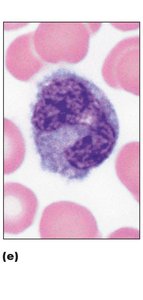
Monocytes: Largest WBCs, U- or kidney-shaped nucleus, differentiate into macrophages in tissues.
Leukopoiesis (White Blood Cell Production)
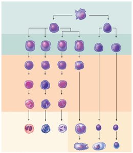
Leukopoiesis is the production of WBCs, stimulated by interleukins and colony-stimulating factors (CSFs). All leukocytes originate from hematopoietic stem cells (hemocytoblasts). Granulocytes and monocytes derive from myeloid stem cells, while lymphocytes derive from lymphoid stem cells.
Leukocyte Disorders
Leukopenia: Abnormally low WBC count, often drug-induced.
Leukemia: Cancerous overproduction of abnormal WBCs, crowding out other blood cell lines, leading to anemia and bleeding.
Infectious Mononucleosis: Viral disease (Epstein-Barr virus) causing high numbers of atypical agranulocytes.
Platelets
Structure and Function
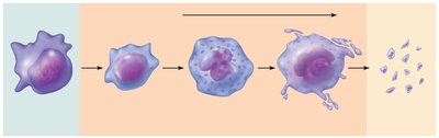
Platelets are cytoplasmic fragments of megakaryocytes, containing granules with chemicals essential for clotting (serotonin, Ca2+, enzymes, ADP, PDGF). They form temporary plugs to seal breaks in blood vessels and are regulated by thrombopoietin.
Hemostasis (Prevention of Blood Loss)
Overview
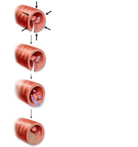
Hemostasis is a fast series of reactions to stop bleeding, involving three main steps:
Vascular spasm: Vasoconstriction of damaged blood vessel.
Platelet plug formation: Platelets adhere to exposed collagen fibers and release chemicals to attract more platelets.
Coagulation: Reinforcement of the platelet plug with fibrin threads, transforming blood from liquid to gel.
Coagulation Pathways
Coagulation involves a cascade of clotting factors, leading to the formation of a fibrin mesh that traps blood cells and seals the injury. There are intrinsic and extrinsic pathways, both converging on the activation of factor X to form prothrombin activator, which converts prothrombin to thrombin. Thrombin then converts fibrinogen to fibrin, forming the clot.
Clot Retraction and Vessel Repair
Clot retraction stabilizes the clot by contracting platelets, pulling the wound edges together. Platelet-derived growth factor (PDGF) and vascular endothelial growth factor (VEGF) stimulate repair of the vessel wall and endothelium.
Fibrinolysis
Fibrinolysis removes unneeded clots after healing. Plasminogen is converted to plasmin, which digests fibrin and dissolves the clot.
Disorders of Hemostasis
Thromboembolic disorders: Undesirable clot formation (thrombus, embolus, embolism).
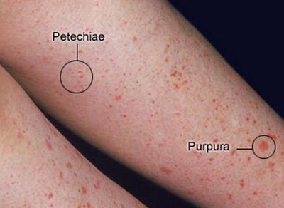
Bleeding disorders: Abnormalities preventing normal clot formation (e.g., thrombocytopenia, hemophilia).
Human Blood Groups and Transfusions
Blood Groups
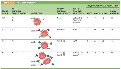
Red blood cell membranes have glycoprotein antigens (agglutinogens) that determine blood groups. The ABO and Rh systems are the most clinically significant. Mismatched transfusions can cause agglutination and hemolysis, leading to serious complications.
Blood Group | RBC Antigens | Plasma Antibodies | Blood That Can Be Received |
|---|---|---|---|
AB | A, B | None | A, B, AB, O (universal recipient) |
B | B | Anti-A | B, O |
A | A | Anti-B | A, O |
O | None | Anti-A, Anti-B | O (universal donor) |
Transfusion Reactions
Transfusion reactions occur if mismatched blood is infused, resulting in agglutination, hemolysis, and possible renal failure. Symptoms include fever, chills, low blood pressure, rapid heartbeat, nausea, and vomiting. Treatment focuses on preventing kidney damage.
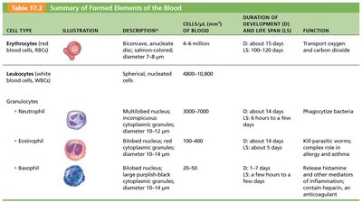
Summary Table: Formed Elements of the Blood
Cell Type | Description | Cells/μL | Duration of Development & Life Span | Function |
|---|---|---|---|---|
Erythrocytes | Biconcave, anucleate discs | 4–6 million | Dev: ~15 days; LS: 100–120 days | Transport oxygen and carbon dioxide |
Leukocytes | Spherical, nucleated cells | 4,800–10,800 | Varies | Defense against disease |
Neutrophils | Multilobed nucleus, pale granules | 3,000–7,000 | Dev: ~14 days; LS: 6 hours–few days | Phagocytize bacteria |
Eosinophils | Bilobed nucleus, red granules | 100–400 | Dev: ~14 days; LS: ~5 days | Kill parasitic worms; role in allergy/asthma |
Basophils | Lobed nucleus, large blue granules | 20–50 | Dev: 1–7 days; LS: few hours–few days | Release histamine, contain heparin |
Lymphocytes | Spherical/indented nucleus | 1,500–3,000 | Dev: days–weeks; LS: hours–years | Mount immune response |
Monocytes | U/kidney-shaped nucleus | 100–700 | Dev: 2–3 days; LS: months | Phagocytosis; develop into macrophages |
Platelets | Discoid cytoplasmic fragments | 150,000–400,000 | Dev: 4–5 days; LS: 5–10 days | Seal small tears in blood vessels; clotting |