 Back
BackBlood Composition and Immune System: Structure, Function, and Communication
Study Guide - Smart Notes
 Tailored notes based on your materials, expanded with key definitions, examples, and context.
Blood Composition
Plasma Components
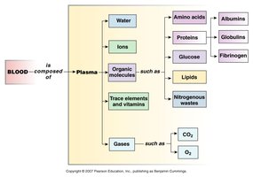
Blood is composed of plasma and cellular elements. Plasma is the liquid portion of blood and serves as a transport medium for nutrients, waste products, and proteins. It contains water, ions, organic molecules, trace elements, vitamins, and gases.
Water: The main solvent in plasma, facilitating transport and chemical reactions.
Ions: Electrolytes such as sodium, potassium, calcium, and chloride help maintain osmotic balance and are essential for cellular function.
Organic Molecules: Includes amino acids, proteins (albumins, globulins, fibrinogen), glucose, lipids, and nitrogenous wastes.
Trace Elements and Vitamins: Required for metabolic processes and enzyme function.
Gases: Oxygen (O2) and carbon dioxide (CO2) are transported in plasma.
Cellular Elements of Blood
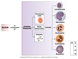
The cellular elements of blood include red blood cells, white blood cells, and platelets. Each plays a distinct role in physiological processes such as oxygen transport, immunity, and clotting.
Red Blood Cells (Erythrocytes): Transport oxygen and carbon dioxide.
White Blood Cells (Leukocytes): Defend against infection and foreign substances.
Platelets: Essential for blood clotting and wound repair.
White Blood Cell Types: Lymphocytes, monocytes, neutrophils, eosinophils, basophils.
Plasma Proteins
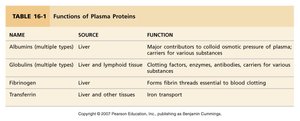
Plasma proteins are synthesized mainly in the liver and perform various functions including maintaining osmotic pressure, transport, immunity, and clotting.
Albumins: Major contributors to colloid osmotic pressure; carriers for various substances.
Globulins: Include clotting factors, enzymes, antibodies, and carriers.
Fibrinogen: Forms fibrin threads essential to blood clotting.
Transferrin: Iron transport protein.
Name | Source | Function |
|---|---|---|
Albumins | Liver | Colloid osmotic pressure, carrier for substances |
Globulins | Liver, lymphoid tissue | Clotting, enzymes, antibodies, carriers |
Fibrinogen | Liver | Blood clotting |
Transferrin | Liver, other tissues | Iron transport |
The Immune System
Key Cells of the Immune System
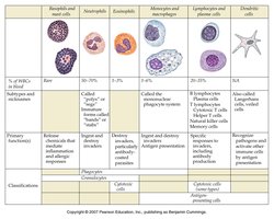
The immune system consists of various cell types, each with specialized functions in defense, recognition, and response to pathogens.
Granulocytes: Include neutrophils, eosinophils, and basophils; involved in phagocytosis, allergic reactions, and inflammation.
Mononuclear Phagocyte System: Monocytes mature into macrophages, which are phagocytic and act as antigen-presenting cells (APCs).
Lymphocytes: Responsible for acquired immunity; include B cells, T cells, and natural killer cells.
Dendritic Cells: Activate lymphocytes and present antigens.
Cell Type | Primary Function | Classification |
|---|---|---|
Neutrophils | Phagocytosis, cytokine release | Granulocyte, phagocyte |
Eosinophils | Destroy invaders, antibody-mediated | Granulocyte, cytotoxic |
Basophils/Mast Cells | Release chemicals for inflammation | Granulocyte |
Monocytes/Macrophages | Phagocytosis, antigen presentation | Phagocyte, APC |
Lymphocytes/Plasma Cells | Specific responses to invaders | B, T, NK cells |
Dendritic Cells | Antigen presentation | APC |
Immune System Functions
The immune system protects the body from pathogens, removes dead or damaged cells, and recognizes and eliminates abnormal cells.
Protection: Against parasites, bacteria, viruses, and foreign molecules.
Removal: Of dead or damaged cells.
Recognition: Of abnormal cells for removal.
Steps in an Immune Response
The immune response is a coordinated process involving detection, communication, recruitment, and destruction of invaders.
Detection: Identify invader or foreign cells.
Communication: Alarm and recruit immune cells.
Coordination: Organize response among participants.
Suppression/Destruction: Eliminate invader.
Cytokine Signaling and T Cell Activation
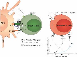
Cytokines are signaling molecules that mediate communication between immune cells. Interleukin-2 (IL-2) is a key cytokine released by helper T cells, promoting proliferation and survival via autocrine signaling.
Autocrine Communication: IL-2 binds to its own receptors on the T cell, activating intracellular pathways (JAK-STAT, MAPK/Erk, Pi3K/Akt).
Positive Feedback: Antigen-activated T cells produce IL-2, stimulating further proliferation.
Antigen Presentation
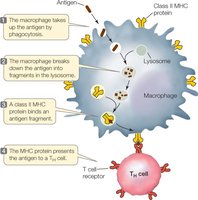
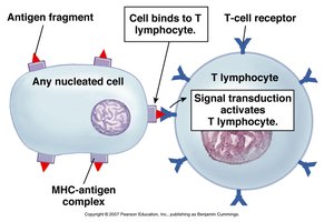
Antigen-presenting cells (APCs) such as macrophages ingest foreign antigens, process them, and present peptide fragments on MHC molecules to T cells, initiating immune activation.
Phagocytosis: APCs ingest and break down antigens.
MHC Presentation: Peptide fragments are displayed on MHC molecules.
T Cell Activation: T cell receptors recognize the antigen-MHC complex, triggering activation.
Inflammatory Response
Inflammation is a protective response involving immune cell recruitment, barrier formation, and tissue repair. Chemical mediators such as histamines, interleukins, bradykinin, and complement proteins play key roles.
Histamines: Released from mast cells, cause vasodilation and increased permeability.
Interleukins: Promote fever and acute-phase protein production.
Bradykinin: Induces pain and swelling.
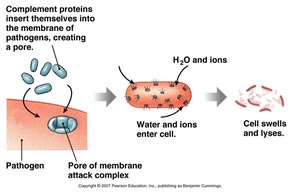
Complement Cascade: Forms membrane attack complex, lysing pathogens.
Chemicals of the Immune Response
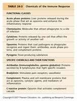
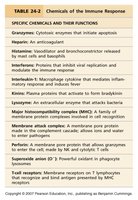
Various chemicals mediate immune responses, including acute phase proteins, chemotaxins, opsonins, pyrogens, antibodies, cytokines, and complement proteins.
Class | Function |
|---|---|
Acute phase proteins | Enhance inflammatory response |
Chemotaxins | Attract phagocytes |
Opsonins | Tag pathogens for phagocytosis |
Pyrogens | Induce fever |
Antibodies | Bind antigens, neutralize pathogens |
Cytokines | Regulate immune cell activity |
Complement proteins | Lyse pathogens, enhance phagocytosis |
Histamine Release and Action
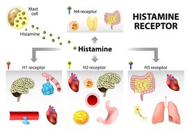
Histamine is released by mast cells during allergic reactions or injury, acting locally via paracrine signaling. It binds to histamine receptors (H1, H2, H3, H4) on target cells, causing vasodilation, increased permeability, smooth muscle contraction, and immune cell recruitment.
Vasodilation: Increases blood flow, causing redness and warmth.
Capillary Permeability: Leads to swelling (edema).
Smooth Muscle Contraction: Causes constriction in airways or gut.
Immune Cell Recruitment: Attracts more white blood cells to the site.
Immune Response to Bacterial Invasion
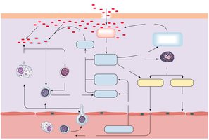
When bacteria enter the extracellular fluid, the immune system responds by activating phagocytes, releasing opsonins and acute phase proteins, and recruiting leukocytes. Complement proteins form membrane attack complexes to lyse bacteria.
Opsonins: Coat bacteria for easier phagocytosis.
Acute Phase Proteins: Enhance immune response.
Complement Proteins: Lyse bacteria via membrane attack complex.
Antibodies: Bind and neutralize bacteria.
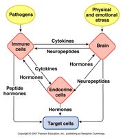
Neuro-Endocrine-Immune Interaction
The nervous, endocrine, and immune systems interact through signaling molecules such as cytokines, hormones, and neuropeptides. Physical and emotional stress can influence immune responses, and immune cells can affect brain and endocrine function.
Cytokines: Mediate communication between immune and brain cells.
Hormones: Influence immune cell activity.
Neuropeptides: Affect both immune and endocrine responses.
Summary Table: Immune Cell Groups
Morphological Group | Functional Group | Examples |
|---|---|---|
Granulocytes | Phagocytes, cytotoxic cells, APCs | Neutrophils, eosinophils, basophils |
Mononuclear phagocyte system | Phagocytes, APCs | Monocytes, macrophages |
Lymphocytes | Acquired immunity | B cells, T cells, NK cells |
Dendritic cells | Activate lymphocytes | Dendritic cells |
Key Equations
Osmotic Pressure Equation:
Where is osmotic pressure, is the van't Hoff factor, is molarity, is the gas constant, and is temperature.
Complement Activation:
Antigen Presentation:
Additional info: Academic context was added to clarify immune cell functions, chemical mediators, and the steps of immune response, as well as to provide self-contained explanations suitable for exam preparation.
