 Back
BackBlood: Composition, Function, and Hemostasis (Chapter 17 Study Notes)
Study Guide - Smart Notes
 Tailored notes based on your materials, expanded with key definitions, examples, and context.
Blood: Composition and Physical Characteristics
Overview of Blood
Blood is the only fluid tissue in the human body, consisting of formed elements suspended in plasma. It plays a critical role in transportation, regulation, and protection within the body.
Formed elements: Erythrocytes (red blood cells, RBCs), leukocytes (white blood cells, WBCs), and platelets.
Physical characteristics: Color ranges from scarlet (oxygen-rich) to dark red (oxygen-poor); more dense and viscous than water; pH 7.35–7.45; constitutes ~8% of body weight (5–6 L in males, 4–5 L in females).
Blood Functions
Distribution: Transports oxygen, nutrients, metabolic wastes, and hormones.
Regulation: Maintains body temperature, pH (via plasma proteins and bicarbonate), and adequate fluid volume.
Protection: Platelets and plasma proteins protect against blood loss; antibodies, complement, and WBCs protect against infection.
Blood Plasma
Composition and Function
Blood plasma is a straw-colored liquid making up about 55% of blood volume. It is 90% water and contains many solutes, including plasma proteins, electrolytes, nutrients, gases, and waste products.
Plasma proteins: Produced mainly by the liver (except gamma globulins, which are antibodies). Major protein is albumin (60%), which acts as a carrier, buffer, and major osmotic protein.
Osmotic ions: Sodium (Na+) is the major osmotic ion in plasma.
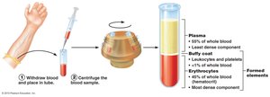
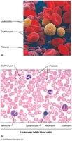
Formed Elements of Blood
Types and Characteristics
The formed elements include erythrocytes, leukocytes, and platelets. Leukocytes are the only complete cells, as erythrocytes lack nuclei and platelets are cell fragments. Most formed elements are short-lived and are constantly replaced by the bone marrow.
Erythrocytes (Red Blood Cells)
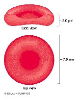
Structure and Function
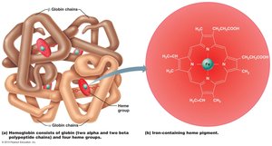
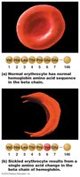
Erythrocytes are biconcave discs (~7.5 μm diameter) lacking nuclei and most organelles. Their primary function is to transport oxygen from the lungs to tissues and carry 20% of carbon dioxide back to the lungs.
Specialized features: Large surface area-to-volume ratio, >97% hemoglobin content, no mitochondria (ATP generated anaerobically).
Hemoglobin (Hb): Each molecule consists of four polypeptide chains (2α, 2β) and four heme groups, each containing an iron atom that binds oxygen reversibly.
Oxyhemoglobin: Oxygen-bound form; deoxyhemoglobin: oxygen released form; carbaminohemoglobin: CO2 bound to globin.
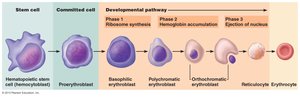
Life Cycle of Erythrocytes
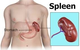
Erythrocytes are produced in the red bone marrow through erythropoiesis, a process regulated by erythropoietin (EPO). Their lifespan is 100–120 days, after which they are removed by the spleen.
Hematopoiesis: General production of formed elements; occurs in red bone marrow.
Erythropoiesis: Production of RBCs from hematopoietic stem cells (hemocytoblasts) through several developmental stages, including reticulocytes (immature RBCs).
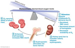
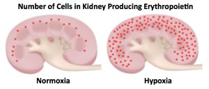
Regulation of Erythropoiesis
The balance between RBC production and destruction is tightly regulated. Erythropoietin (EPO), primarily produced by the kidneys, stimulates RBC production in response to hypoxia (low oxygen levels).
Stimuli for EPO release: Hemorrhage, high altitude, increased tissue demand for oxygen.
Dietary requirements: Iron, vitamin B12, and folic acid are essential for erythropoiesis.
Destruction of Erythrocytes
Old erythrocytes become rigid and fragile, and are phagocytized in the spleen. Iron is recycled, heme is degraded to bilirubin (excreted in bile), and globin is broken down to amino acids.
Disorders of Erythrocytes
Anemia and Polycythemia
Anemia: Reduced oxygen-carrying capacity due to insufficient RBCs, blood loss, or increased destruction. Symptoms include fatigue, pallor, and shortness of breath.
Polycythemia: Excess RBCs increase blood viscosity, risking clotting and impaired circulation. Can be primary (polycythemia vera), secondary (due to hypoxia), or artificial (blood doping).
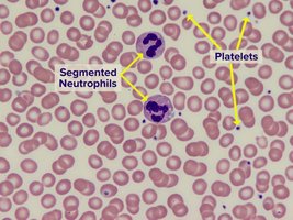
Platelets and Hemostasis
Platelet Structure and Function
Platelets are cytoplasmic fragments of megakaryocytes, essential for blood clotting. They contain granules with clotting factors and enzymes, are anucleate, and have a lifespan of ~10 days. Platelet production is regulated by thrombopoietin.

Hemostasis: Stopping Blood Loss
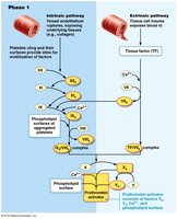
Hemostasis is the process that prevents blood loss after vessel injury, involving three major steps:
Vascular spasm: Vasoconstriction of damaged vessel to reduce blood flow.
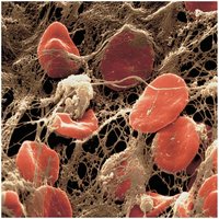
Platelet plug formation: Platelets adhere to exposed collagen (via von Willebrand factor), become activated, and aggregate to form a temporary plug.
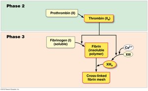
Coagulation: Reinforcement of the platelet plug with fibrin threads, forming a stable clot.
Coagulation Pathways
Coagulation involves a cascade of reactions leading to the conversion of fibrinogen to fibrin. There are two pathways to prothrombin activator (PA):
Intrinsic pathway: Initiated by damage to the vessel wall and exposure to collagen; slower.
Extrinsic pathway: Triggered by tissue factor released from damaged tissues; faster.
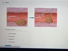
Clot Retraction and Repair
After coagulation, the clot contracts (clot retraction) to bring wound edges closer together. Platelet-derived growth factor (PDGF) and vascular endothelial growth factor (VEGF) stimulate tissue repair.
Blood Types and Transfusion Reactions
ABO and Rh Blood Groups
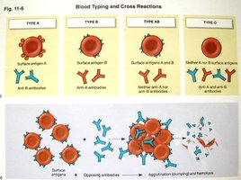
Blood types are determined by the presence or absence of antigens (A, B, Rh) on the surface of erythrocytes. The ABO system includes four main types: A, B, AB, and O. The Rh factor is another important antigen (positive or negative).
Type A: Surface antigen A; anti-B antibodies in plasma.
Type B: Surface antigen B; anti-A antibodies in plasma.
Type AB: Both A and B antigens; no anti-A or anti-B antibodies (universal recipient).
Type O: No A or B antigens; both anti-A and anti-B antibodies (universal donor).
Transfusion reactions occur if incompatible blood is transfused, leading to agglutination and hemolysis of donor cells.
Summary Table: ABO Blood Types
Blood Type | Surface Antigens | Plasma Antibodies | Can Receive From | Can Donate To |
|---|---|---|---|---|
A | A | Anti-B | A, O | A, AB |
B | B | Anti-A | B, O | B, AB |
AB | A, B | None | A, B, AB, O | AB |
O | None | Anti-A, Anti-B | O | A, B, AB, O |
Additional info: The Rh factor is especially important in pregnancy (hemolytic disease of the newborn) and transfusion compatibility. Major anticoagulants in the body include antithrombin and heparin, which prevent unwanted clotting.
