 Back
BackBlood: Composition, Functions, and Clinical Relevance
Study Guide - Smart Notes
 Tailored notes based on your materials, expanded with key definitions, examples, and context.
The Blood
Overview: Composition of Blood
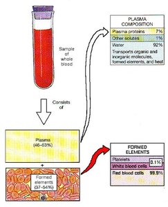
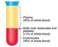
Blood is a specialized liquid connective tissue essential for the transport of substances throughout the body. It consists of two main components: the formed elements (cells and platelets) and the plasma (fluid matrix). Blood is denser and more viscous than water due to dissolved ions, organic molecules, plasma proteins, and blood cells. The average temperature is 38°C, and the pH is tightly regulated between 7.35 and 7.45. Blood volume varies by sex, with females averaging 4-5 L and males 5-6 L.
Formed elements: Living blood cells (erythrocytes, leukocytes) and platelets (thrombocytes).
Plasma: Fluid matrix containing water, proteins, and solutes.
Hematocrit: Percentage of blood volume occupied by formed elements (~45%).
Functions of Blood
Blood performs three primary functions: transport, regulation, and protection.
Transport: Delivers oxygen, nutrients, and hormones; removes carbon dioxide and metabolic wastes.
Regulation: Maintains body temperature, pH, fluid volume, and composition of interstitial fluid/lymph.
Protection: Facilitates inflammation and repair, prevents blood loss (hemostasis), and defends against infection.
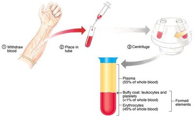
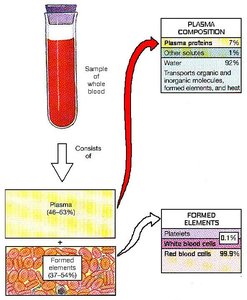
Components of Blood
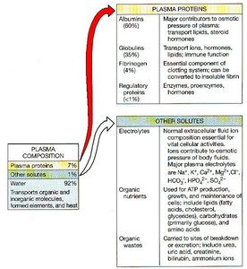
Plasma
Plasma is the straw-colored liquid portion of blood, making up about 55% of its volume. It is composed of 92% water, 7% proteins, and 1% other solutes.
Proteins: Albumin (60%), globulins (35%), fibrinogen (4%), and regulatory proteins (1%).
Other solutes: Waste products, nutrients, electrolytes, enzymes, hormones, and gases (O2, CO2, N2).
Component | Percentage | Main Functions |
|---|---|---|
Water | 92% | Solvent for transport |
Proteins | 7% | Osmotic balance, clotting, immunity |
Other solutes | 1% | Electrolytes, nutrients, wastes |
Formed Elements
The formed elements constitute about 45% of blood volume and include erythrocytes, leukocytes, and platelets.
Erythrocytes (RBCs): >99% of formed elements; transport O2 and CO2.
Leukocytes (WBCs): <1%; immune defense.
Thrombocytes (Platelets): <1%; blood clotting.
Formed Element | Percentage | Function |
|---|---|---|
Red blood cells | 99.9% | Oxygen and carbon dioxide transport |
White blood cells | 0.1% | Immune defense |
Platelets | 0.1% | Clotting |
Hematopoiesis
Blood Cell Formation
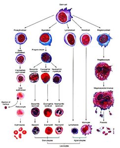
All blood cells originate from pluripotent hematopoietic stem cells in the red bone marrow. These stem cells differentiate into five types of precursor cells, which further develop into RBCs, WBCs, and megakaryocytes (which produce platelets).
Production of Erythrocytes (Erythropoiesis)
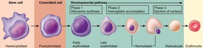
Erythropoiesis is the process of RBC production, regulated by erythropoietin (EPO) from the kidneys. It involves three phases: ribosome production, hemoglobin synthesis, and ejection of the nucleus. RBCs are released as reticulocytes and mature in the bloodstream.
Regulation: Negative feedback based on O2 levels; hypoxia increases EPO production.
Numbers: Males: 5.4 million RBCs/ml; Females: 4.8 million RBCs/ml.
RBC Production - Anemia
Anemia is characterized by reduced O2 carrying capacity of blood. Causes include insufficient RBCs, hemolytic anemia, destruction of bone marrow, decreased hemoglobin content, iron deficiency, and vitamin B12 deficiency. Hereditary forms include thalassemias and sickle cell anemia.
Thalassemias: Reduced or absent hemoglobin synthesis; fragile RBCs.
Sickle Cell Anemia: Mutation in hemoglobin; sickled cells block capillaries and reduce O2 transport.
Hematocrit (Hct)
Hematocrit is the percentage of blood that is RBCs. Normal values: Males 40-54% (47%), Females 38-46% (42%). It indicates RBC production and hydration status. Abnormal values may result from altitude, blood doping, anemia, hemorrhage, malaria, cancer, chemotherapy, radiation, or drugs.
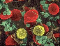
Erythrocytes (Red Blood Cells)
Anatomy and Physiology
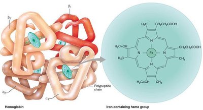
Erythrocytes are biconcave disks, 8 µm in diameter, lacking a nucleus and most organelles. They are filled with hemoglobin, which constitutes 33% of cell weight and is responsible for O2 transport.
Biconcave shape: Increases surface area for gas diffusion and flexibility for capillary passage.
Hemoglobin: Composed of 4 globin chains (2 α, 2 β) and 4 heme groups, each with an iron ion (Fe2+) that binds O2.
RBCs: Carry ~1 billion O2 molecules per cell; also transport ~25% of CO2.
RBC Life Span
RBCs have a life span of 100-120 days. They cannot repair damage due to the absence of a nucleus and ribosomes. Old RBCs are destroyed in the spleen, liver, and bone marrow. Breakdown products are recycled: iron is reused, and the non-iron portion is converted to bilirubin, contributing to the color of bruises, urine, and feces.
Leukocytes (White Blood Cells)
Granular Leukocytes
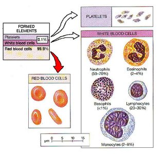
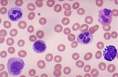
Granular leukocytes include neutrophils, eosinophils, and basophils. Each type has distinct anatomical and physiological characteristics.
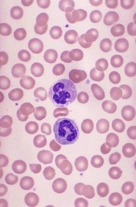
Neutrophils: 60-70% of WBCs; phagocytize bacteria, release lysozyme, oxidants, and defensins.
Eosinophils: 2-4% of WBCs; combat viruses and parasites, produce histamine, phagocytize antigen-antibody complexes.
Basophils: 0.5-1% of WBCs; release heparin, histamine, serotonin; involved in allergic reactions.

Agranular Leukocytes
Agranular leukocytes include lymphocytes and monocytes. Lymphocytes are central to immune responses, while monocytes differentiate into macrophages for cleanup.
Lymphocytes: 20-25% of WBCs; B-cells produce antibodies, T-cells attack infected cells.
Monocytes: 3-8% of WBCs; become macrophages, clean up debris and microbes.
Leukocyte Type | Percentage | Function |
|---|---|---|
Neutrophils | 60-70% | Phagocytosis of bacteria |
Lymphocytes | 20-25% | Immune response |
Monocytes | 3-8% | Cleanup, macrophage formation |
Eosinophils | 2-4% | Combat parasites, viruses |
Basophils | 0.5-1% | Allergic reactions |
Leukocyte Life Span and Number
Leukocyte life span varies with activity. Most live days to months, but during infection, some may only survive hours. The normal WBC count is 5,000-10,000/mm3 blood, with a RBC/WBC ratio of 700:1. Abnormalities include leukopenia (decreased numbers), leukocytosis (increased numbers), and leukemia/lymphoma (cancerous proliferation).
Thrombocytes (Platelets)
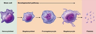
Development and Anatomy
Platelets are cytoplasmic fragments shed from megakaryocytes. They lack a nucleus, are disc-shaped, and measure 2-4 µm in diameter. Normal count is 250,000-400,000/mm3.
Physiology
Platelets have a short life span (5-9 days) and help plug small holes in blood vessels. Their granules contain regulatory factors for coagulation, inflammation, and immune defenses.
Hemostasis
Mechanisms to Stop Bleeding
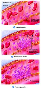
Hemostasis involves three mechanisms: vascular spasm, platelet plug formation, and coagulation.
Vascular spasm: Immediate constriction of damaged blood vessels.
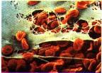
Platelet plug formation: Platelets adhere to exposed collagen, activate, aggregate, and block blood loss in small vessels.
Coagulation: Formation of a gel (clot) in plasma that traps formed elements. Fibrinogen is converted to fibrin, forming a mesh.
Coagulation is a positive feedback cascade involving numbered factors and calcium ions (Ca2+).
Blood Types
ABO Blood Typing
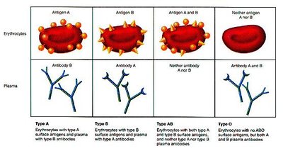
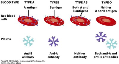
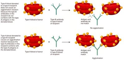
Blood types are determined by the presence of agglutinogens (antigens) A and B on RBC surfaces. Agglutinins (antibodies) are produced against antigens not present in one's blood. Blood type AB is the universal recipient; type O is the universal donor.
Type A: Antigen A, anti-B antibody
Type B: Antigen B, anti-A antibody
Type AB: Antigens A and B, no antibodies
Type O: No antigens, both anti-A and anti-B antibodies
Blood Type | Antigen(s) | Antibody(ies) | Transfusion Compatibility |
|---|---|---|---|
A | A | Anti-B | Can receive A, O |
B | B | Anti-A | Can receive B, O |
AB | A, B | None | Can receive A, B, AB, O |
O | None | Anti-A, Anti-B | Can receive O only |
Rh Typing
Rh typing is based on the presence of Rh antigen. Individuals with Rh antigen are Rh+, those without are Rh-. Rh agglutinins are only produced after exposure to Rh antigen. Rh sensitivity can cause hemolytic disease of the newborn (erythroblastosis fetalis).
Additional info: The notes above expand on brief points with academic context, definitions, and examples to ensure completeness and clarity for exam preparation.
