 Back
BackBlood: Structure, Components, and Typing – ANP College Study Guide
Study Guide - Smart Notes
 Tailored notes based on your materials, expanded with key definitions, examples, and context.
Blood: Structure and Function
Overview of Blood as a Connective Tissue
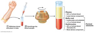
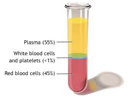
Blood is a specialized connective tissue composed of formed elements (cells and cell fragments) suspended in an intercellular material called plasma. It plays a critical role in transport, regulation, and protection within the human body.
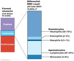
Formed elements: Erythrocytes (red blood cells), leukocytes (white blood cells), and platelets (cell fragments).
Plasma: The liquid matrix that carries solutes and formed elements.
Physical Characteristics of Blood
Volume: Adult males: 5–6 liters; adult females: 4–5 liters.
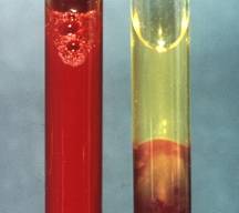
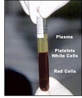
Hematocrit: The percentage of blood volume occupied by red blood cells.
Adult males: 47% ± 5%
Adult females: 42% ± 5%
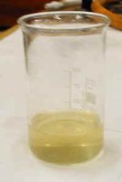
Blood Plasma
Characteristics and Composition
Plasma is the extracellular material of blood, serving as the medium for transport of nutrients, wastes, and other molecules.
pH: 7.35–7.45 (slightly alkaline)
Water content: 90%
Solutes (10%):
Plasma proteins: Albumin, antibodies, coagulation proteins
Nutrients: Proteins, carbohydrates, lipids, vitamins
Hormones
Wastes: Urea, uric acid, creatinine
Dissolved gases: CO2, O2
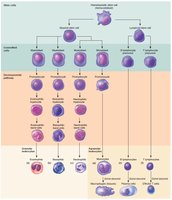
Formed Elements of Blood
Types and Functions
The formed elements include erythrocytes, leukocytes, and platelets, each with distinct structure and function.
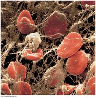
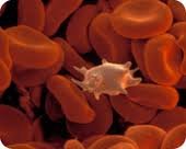
Erythrocytes (Red Blood Cells, RBCs):
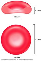
Anucleate cells specialized for oxygen and carbon dioxide transport.
Contain hemoglobin, which binds O2 (oxyhemoglobin) and CO2 (carbaminohemoglobin).
Biconcave shape, diameter ~7.5 μm.
Normal count: 4–6 × 10⁶ cells/mm3 blood.
Leukocytes (White Blood Cells, WBCs):
Nucleated cells providing immunity.
Normal count: 4,800–10,800 cells/mm3 blood.
Five types, divided into granulocytes and agranulocytes.
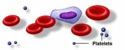
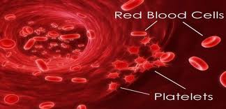
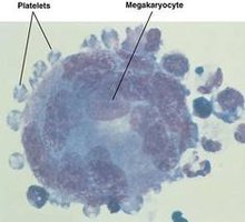
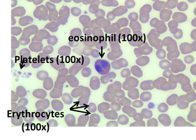
Platelets (Thrombocytes):
Cell fragments derived from megakaryocytes.
Contain granules, no nuclei or organelles.
Function in hemostasis (prevention of blood loss).
Normal count: 150,000–400,000 cells/mm3 blood.
Red Blood Cells (Erythrocytes)
Lack nuclei and organelles.
Contain hemoglobin for gas transport.
Biconcave shape increases surface area for gas exchange.
Diameter: 7.5 μm; thickness: 2.0 μm.

Leukocytes: Classification and Identification
Leukocytes are divided into two main classes:
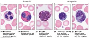
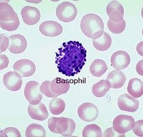
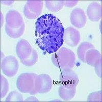
Granulocytes: Neutrophils, eosinophils, basophils
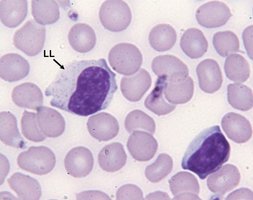
Agranulocytes: Lymphocytes, monocytes
Mnemonic for Leukocyte Abundance
Never Let Monkeys Eat Bananas (Neutrophils > Lymphocytes > Monocytes > Eosinophils > Basophils)
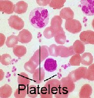
Platelets (Thrombocytes)
Fragments from megakaryocytes.
Contain granules, lack nuclei.
Essential for blood clotting (hemostasis).
Blood Smear and Staining
Wright's Stain: Identification of Formed Elements
Wright's stain is a differential stain used to distinguish blood cells.
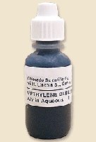
Methylene Blue: Basic dye, stains acidic components deep blue/purple (basophilic structures).
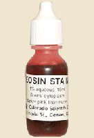
Eosin: Acidic dye, stains basic components red/deep pink/orange (acidophilic structures).
Eosin/Methylene Blue Complex: Stains neutral substances lilac.
Blood Smear Identification
When blood is smeared, dried, and stained, individual formed elements can be distinguished, including types of WBCs. 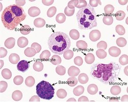
Differential White Blood Cell Count
Purpose and Clinical Significance
A differential WBC count determines the relative percentage of each type of leukocyte in peripheral blood.
Used to detect diseases: acute/chronic infection, allergy, parasitic diseases, anemia, HIV, etc.
Each leukocyte type has a normal range in peripheral blood.
Leukocyte Types: Structure and Function
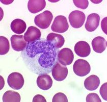
Granulocytes
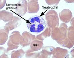
Neutrophils: 50–70% of WBCs; 9–16 μm; 2–5 nuclear lobes; pale lavender granules.
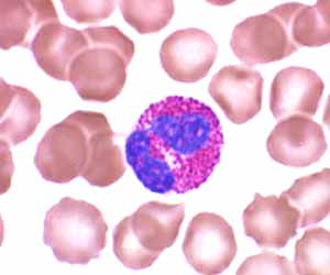
Eosinophils: 2–4% of WBCs; 10–14 μm; bi-lobed nucleus; bright reddish/orange/pink granules.
Basophils: 0–1% of WBCs; 8–10 μm; unsegmented/bilobed nucleus; deep blue/purple granules.
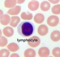
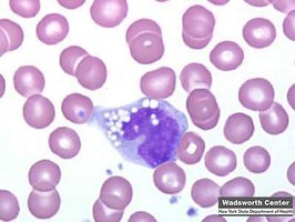
Agranulocytes
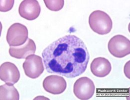
Lymphocytes: 25–45% of WBCs; 5–17 μm; round/slightly oval nucleus; clear blue cytoplasm.
Monocytes: 3–8% of WBCs; 14–24 μm; horseshoe/kidney-shaped nucleus; blue-gray cytoplasm with vacuoles.
Blood Typing
ABO and Rh Blood Group Systems
Blood typing is based on the presence of specific agglutinogens (antigens) on RBC surfaces.
ABO System: Types A, B, AB, O
Rh System: Rh+ (D antigen present), Rh- (D antigen absent)
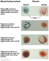
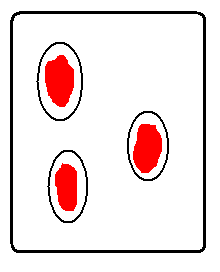
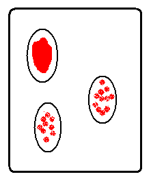
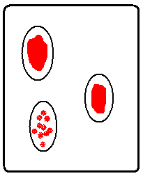
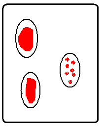
Blood Type Determination
Mix blood sample with anti-A, anti-B, and anti-D agglutinins.
Agglutination indicates presence of corresponding agglutinogen.
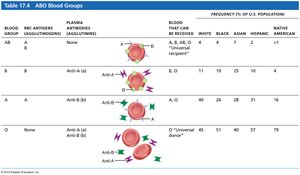

Blood Group Compatibility
Blood Group | RBC Antigens | Plasma Antibodies | Blood Received |
|---|---|---|---|
AB | A, B | None | A, B, AB, O |
A | A | Anti-B | A, O |
B | B | Anti-A | B, O |
O | None | Anti-A, Anti-B | O |
Agglutination vs. Coagulation
Agglutination: Clumping reaction when agglutinins (antibodies) bind to agglutinogens (antigens) on RBCs.
Coagulation: Formation of a blood clot by platelets and clotting factors.
Safety Precautions in Blood Labs
Wear gloves and goggles.
Do not test your own blood; use provided samples.
Materials contacting blood must be placed in bleach solution.
Avoid spills; if a spill occurs, cover with bleach and notify instructor.
Summary Table: Formed Elements and Their Functions
Formed Element | Function | Normal Range |
|---|---|---|
Erythrocytes | Transport O2 and CO2 | 4–6 × 10⁶/mm3 |
Leukocytes | Immunity | 4,800–10,800/mm3 |
Platelets | Hemostasis | 150,000–400,000/mm3 |
Key Equations
Hematocrit Calculation
Hematocrit (%) =
Blood Type Compatibility
Recipient must not have antibodies against donor's RBC antigens.
Additional info:
Blood typing is essential for safe transfusions and organ transplants.
Wright's stain is a standard method for identifying blood cell morphology in clinical labs.
