 Back
BackBlood: Structure, Function, and Cellular Components (Anatomy & Physiology II, Chapter 19)
Study Guide - Smart Notes
 Tailored notes based on your materials, expanded with key definitions, examples, and context.
Blood: Structure and Functions
Overview of Blood Functions
Blood is a vital connective tissue that performs several essential functions in the human body. It transports substances, regulates physiological parameters, restricts fluid losses, defends against pathogens, and stabilizes body temperature.
Transport: Blood carries gases (O2, CO2), nutrients, hormones, and metabolic wastes.
Regulation: Maintains pH and ion balance in interstitial fluids.
Restriction: Prevents fluid loss at injury sites via clotting.
Defense: Transports white blood cells and antibodies to fight infections.
Temperature Stabilization: Absorbs and redistributes heat throughout the body.
Physical Characteristics and Sampling of Blood
Physical Properties
Blood exhibits distinct physical characteristics that are important for its function and clinical assessment.
Temperature: 100.4°F (slightly above body temperature)
Viscosity: 5 times more viscous than water
pH: 7.35–7.45
Volume: Males: 5–6 liters; Females: 4–5 liters
Blood Collection Methods
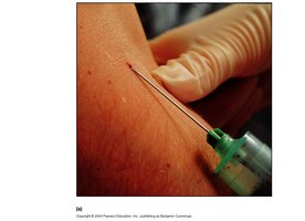
Blood is commonly collected for diagnostic purposes using venipuncture or arterial puncture.
Venipuncture: Most common, taken from the median cubital vein; veins are easier to locate and have lower pressure.
Arterial Puncture: Drawn from radial or brachial artery; used to assess gas exchange efficiency in the lungs.
Composition of Blood
Main Components
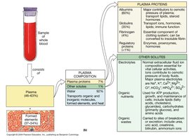
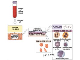
Blood consists of plasma (liquid component) and formed elements (cells and cell fragments).
Plasma: 55–60% of blood; yellowish fluid containing proteins, solutes, and water.
Formed Elements: 40–45% of blood; includes red blood cells (RBCs), white blood cells (WBCs), and platelets.
Plasma Composition
Plasma Proteins: Albumin (60%), Globulins (35%), Fibrinogen (4%), Regulatory proteins (<1%)
Other Solutes: Electrolytes (Na+, K+), organic nutrients, organic wastes
Water: Most abundant component (92%)
Formed Elements of Blood
Red Blood Cells (Erythrocytes)
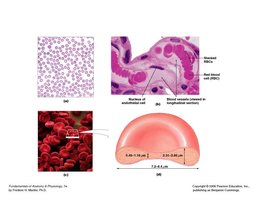
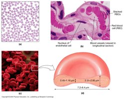
RBCs are specialized for oxygen and carbon dioxide transport. Their unique structure enhances their function.
Shape: Biconcave disk, thin center, thick edge
Features: No nucleus or mitochondria; high surface area for gas exchange; flexible and stackable for capillary passage
Lifespan: Approximately 120 days
Hemoglobin Structure and Function
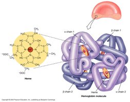
Hemoglobin is the protein responsible for oxygen transport in RBCs. It consists of four polypeptide chains (2 alpha, 2 beta), each with a heme group containing iron.
Oxyhemoglobin (HbO2): Oxygen-bound form; bright red color
Deoxyhemoglobin: Oxygen released
Carbaminohemoglobin: CO2 bound
Each hemoglobin molecule can carry up to four O2 molecules
Abnormal Hemoglobin and Anemia
Iron Deficiency Anemia: Not enough iron to make hemoglobin
Pernicious Anemia: Low RBC production due to lack of vitamin B12 absorption
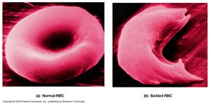
Sickle Cell Anemia: Mutation in beta chain causes RBCs to become stiff and curved
Thalassemia: Inability to produce adequate alpha or beta chains
RBC Recycling and Destruction
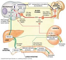
Old or damaged RBCs are broken down by macrophages in the liver and spleen. Hemoglobin is split into heme and globin; iron is recycled, and heme is converted to biliverdin and then bilirubin.
Biliverdin: Green pigment
Bilirubin: Yellow/orange pigment; excreted in bile
Jaundice: Accumulation of bilirubin causes yellow skin
Blood Types and Antigens
Blood Typing
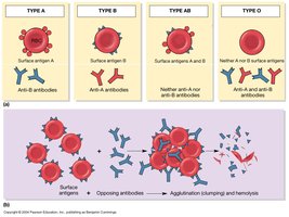
Blood type is determined by the presence or absence of surface antigens (A, B, Rh) on RBCs. Antibodies in plasma react with foreign antigens, causing agglutination.
Type A: Surface antigen A, anti-B antibodies
Type B: Surface antigen B, anti-A antibodies
Type AB: Both antigens, no anti-A/B antibodies (universal recipient)
Type O: No antigens, both anti-A and anti-B antibodies (universal donor)
Rh factor: Rh+ (antigen present), Rh- (antigen absent)
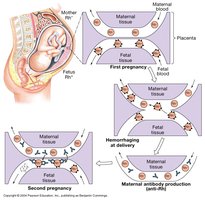
Erythroblastosis Fetalis (Hemolytic Disease of the Newborn)
Occurs when an Rh- mother develops antibodies against Rh+ fetal blood, which can cross the placenta and attack the fetus in subsequent pregnancies.
Origin and Production of Formed Elements
Hematopoiesis
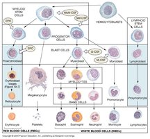
Hematopoiesis is the process of blood cell formation, occurring primarily in red bone marrow. Hemocytoblasts (pluripotent stem cells) differentiate into myeloid and lymphoid stem cells, which give rise to RBCs, WBCs, and platelets.
RBCs: Hemocytoblast → myeloid stem cell → proerythroblast → erythroblast → reticulocyte → erythrocyte
WBCs: Hemocytoblast → myeloid/lymphoid stem cell → progenitor → blast cell → mature leukocyte
Platelets: Hemocytoblast → myeloid stem cell → megakaryocyte → platelets
White Blood Cells (Leukocytes)
Classification and Functions
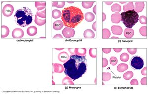
WBCs are divided into granular and agranular types, each with specific roles in immunity.
Granular Leukocytes: Neutrophils, Eosinophils, Basophils
Agranular Leukocytes: Monocytes, Lymphocytes
Key Characteristics
Amoeboid Movement: Enables mobility
Diapedesis: Ability to migrate out of bloodstream
Positive Chemotaxis: Attraction to chemical signals
Phagocytosis: Neutrophils, eosinophils, monocytes engulf pathogens
Types of WBCs
Neutrophils (50–70%): First responders, phagocytize bacteria, form pus
Eosinophils (2–4%): Defend against parasites, involved in allergic responses
Basophils (<1%): Release histamine and heparin, mediate inflammation
Monocytes (2–8%): Become macrophages, present antigens to T-cells
Lymphocytes (20–30%): T-cells (cell-mediated immunity), B-cells (antibody production), NK cells (kill abnormal cells)
Platelets and Hemostasis
Platelet Structure and Function
Platelets are cell fragments essential for blood clotting. They aggregate, adhere, and form plugs at injury sites.
Lifespan: 7–10 days
Properties: Agglutination, adhesiveness, aggregation
Hemostasis: Phases of Blood Clotting
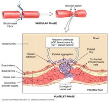
Hemostasis is the process that prevents blood loss after injury, involving three phases:
Vascular Phase: Vessel contracts to reduce blood flow
Platelet Phase: Platelets adhere and form a plug
Coagulation Phase: Cascade of reactions forms a fibrin clot
Blood Clotting Inhibitors and Disorders
Anticoagulants: Antithrombin III, heparin, thrombomodulin, prostacyclin
Hemophilia: Genetic disorder causing inadequate clotting factor production
Embolus: Drifting blood clot; can cause infarction
Thrombus: Clot inside vessel; associated with plaque
Key Definitions
Plasma: Liquid component of blood
Antibodies: Proteins that bind antigens
Hemocytoblasts: Blood stem cells
Hematocrit: Percentage of blood volume occupied by cells
Oxyhemoglobin: Oxygen-bound hemoglobin
Heme molecules: Iron-containing pigment in hemoglobin
Anemia: Reduced oxygen-carrying capacity
Jaundice: Yellow skin due to bilirubin accumulation
Hypoxia: Low oxygen levels in tissues
RhoGam: Medication to prevent Rh antibody formation
Hypovolemic: Low blood volume
Viscosity: Thickness of blood
Macrophage: Phagocytic cell derived from monocytes
Coagulation: Blood clotting process
Embolism: Blockage by a clot
Infarction: Tissue death due to blocked blood supply
Thrombus: Stationary blood clot
Key Equations
Hematocrit Calculation:
Oxygen Carrying Capacity:
